This is an automatically translated article.
The article is professionally consulted by Dr. Nguyen Dinh Hung - Diagnostic Imaging - Department of Diagnostic Imaging - Vinmec Hai Phong International General Hospital.
Upper-abdominal computed tomography with visceral angiography is a technique that uses X-rays of a multi-sequential tomography machine to diagnose, detect, and search for lesions and pathologies of the internal organs. the upper abdominal cavity such as liver, bile, pancreas, stomach, duodenum, spleen, ... and examine the vessels of these organs.
1. Overview of upper abdominal computed tomography scan with visceral vascular examination
Upper-abdominal computed tomography with vascular examination of organs is the use of a system of equipment, multi-segment computed tomography scanners and software to process, reconstruct images, and visualize blood vessels. of the viscera in the upper abdomen including stomach, duodenum, liver, bile, pancreas, spleen, ...
This is a modern technique, allowing simultaneous assessment of the organ's parenchymal status and morphology , pathology of the vessels supplying blood to the viscera, vascular peduncles, and drainage in cases of vascular malformations and tumors.

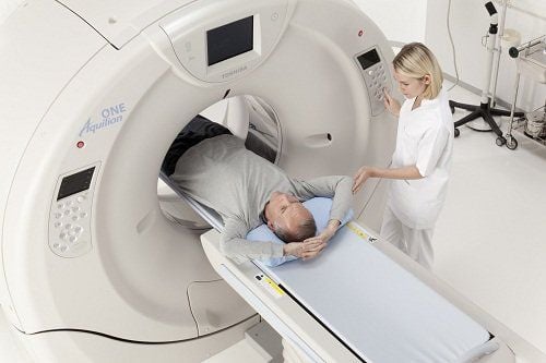
Chụp cắt lớp vi tính tầng trên ổ bụng có khảo sát mạch các tạng là kỹ thuật hiện đại, cho phép bá sĩ chẩn đoán hình ảnh chính xác
2. Indications and contraindications for upper abdominal computed tomography (CT) scan with visceral vascular examination
Upper-abdominal computed tomography with vascular examination of visceral organs is indicated in cases of examination to detect disease and trauma to organs in the upper abdomen, specifically:
● Stomach, duodenum Colon: Trauma, gastrointestinal bleeding, tumor.
● Pancreas: Acute or chronic pancreatitis, pancreatic tumor.
● Spleen: Trauma, tumor.
Biliary tract, gallbladder: Gallstones, gallbladder, biliary tract tumor, gallbladder.
Liver: Trauma, hepatitis, liver abscess, liver tumor.
● Patients before or after organ transplant surgery.
● Other lesions: Subdiaphragmatic abscess, mesenteric tumor.
Visceral vascular examination: Suspect and evaluate thrombosis in the branches of the visceral trunk and superior mesenteric arteries, as well as the portal and hepatic veins in case of tumor lesions. .
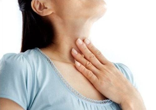
In case the patient is allergic or has a history of allergy to contrast, it is relatively contraindicated and in the case of women in the first weeks of pregnancy should be considered.

Trường hợp bệnh nhân mang thai cần cân nhắc khi chụp cắt lớp vi tính
3. Upper abdominal CT scan procedure with visceral vascular survey
The procedure for taking a CT scan of the upper abdomen with the examination of the visceral pulse includes the following steps:
● Step 1: Place the patient in supine position, raise both arms above the head to avoid image interference. Instruct and ask the patient to hold their breath during the scan to avoid image noise.
● Step 2: Set up an intravenous line to inject iodinated contrast with a dose of 1.5-2 ml/kg body weight. Use a syringe pump for rapid injection with the required injection rate of 3 - 4 ml/s depending on the strength of the blood vessel wall. Depending on the size of the individual change the field of view accordingly and to be able to assess the entire skeletal system, fat, air and soft tissues, change the width of the window.
● Step 3: Computed tomography of the upper abdomen with cross-sectional layers, the thickness of the cut is 5mm, taken before contrast injection to locate the lesion. Assess the lesion composition including fat as well as bleeding and calcification by measuring the density of the suspected lesion. Compare with the density of the lesion area after contrast injection to assess the extent of drug permeability of the lesion more or less.
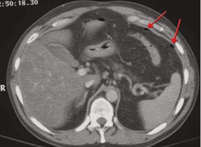
Chụp cắt lớp vi tính tầng trên ổ bụng với các lớp cắt ngang, độ dày lớp cắt là 5mm
● Step 4: After contrast injection, computed tomography of the upper abdomen with cross-sectional layers with an ideal thickness of 2.5 mm, reconstructed layers with thickness of 1mm. Computed tomography in the arterial phase after injection from 25 to 30 seconds to review and evaluate the following conditions: vascular richness of the tumor; perfusion disorders in the parenchyma in solid organs, and at the same time detecting drainage in arteriovenous malformation lesions, visceral trauma causes drugs to leak out into the lumen.
● Step 5: Take scans in the venous phase after contrast injection from 60 to 70 seconds to review and evaluate the following conditions: tumor clearance, organ trauma. Computed tomography (CT) of the upper abdomen with sections in the late phase of 3 - 10 minutes.
● Step 6: The technician recreates images on the scanner and in the imaging chamber, examines the vessels of the viscera and renders the blood vessels in different directions, each organ includes its own arteriovenous system. The image results must ensure no image noise to serve the diagnosis.
All questions need to be answered by a specialist doctor as well as customers wishing to examine and perform an upper abdominal CT scan with an examination of the blood vessels of the organs at Vinmec International General Hospital.
Doctor Nguyen Dinh Hung has over 10 years of experience in the field of diagnostic imaging (Ultrasound, CT, MRI). Trained and practiced on hepatobiliary interventional radiology at Bach Mai Hospital (Intervention under ultrasound guidance, DSA, CT...) and deployed at the Diagnostic Imaging Department of Viet Tiep Hospital Hai Phong. Currently, he is a doctor at the Diagnostic Imaging Department of Vinmec Hai Phong International General Hospital.
Please dial HOTLINE for more information or register for an appointment HERE. Download MyVinmec app to make appointments faster and to manage your bookings easily.
MORE
Contrast in computed tomography (CT Scan) Computed tomography - the gold standard in diagnosing coronary artery disease What is CT? In which cases need contrast injection?