This is an automatically translated article.
The article is professionally consulted by Master, Doctor Nguyen Viet Thu - Doctor of Radiology and Nuclear Medicine - Department of Diagnostic Imaging and Nuclear Medicine - Vinmec Times City International General Hospital.Computed tomography of the lung has high resolution with the aim of capturing images of lung parenchyma with the best image quality, thereby helping doctors accurately diagnose lung parenchymal and interstitial lung disease, avoiding Ignore minor injuries.
1. What is high resolution computed tomography of the lung?
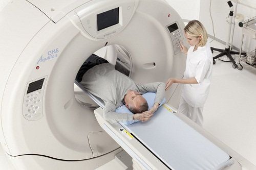
High-resolution computed tomography (CT) scan is a technique that uses X-rays to beam into the lung in slices to obtain high-resolution images of lung parenchyma, helping to diagnose bronchial and interstitial lung diseases.High-resolution lung computed tomography scan is a safe, non-invasive imaging method, but gives high accuracy results. Can diagnose dangerous diseases such as: Pneumonia, lung cancer, determine the status of cancer in other organs with metastasis to the lungs.
2. Indications, contraindications to high-resolution computed tomography of the lung
2.1 Indications of high-resolution computed tomography ● Purpose of finding bronchial lesions such as: bronchiectasis, alveolar dilation, pneumothorax...● Identifying interstitial lung disease.
2.2 Contraindications High resolution lung computed tomography method does not inject contrast, so there is no absolute contraindication. There are only relative contraindications such as:
● Pregnant women, especially in the first 3 months of pregnancy. You should inform your doctor about your pregnancy. In case a woman suspects that she is pregnant, she should take a pregnancy test before deciding to take a scan. When taking pictures for pregnant women, it is necessary to wear a sister shirt to cover and protect the abdomen..
● Have bronchial asthma; atopic allergy to the drug or to other allergens; have kidney disease such as chronic kidney disease, kidney failure...

Phụ nữ mang thai 3 tháng đầu có chống chỉ định tương đối với kỹ thuật này
3. High resolution lung computed tomography scan
3.1 Preparation Performer: Doctor and specialist radiologist.Computer tomography facilities include:
● Computed tomography machine
● Film, printer, image storage system.
Patient
● Patient is clearly explained on how to perform the procedure to coordinate with the radiologist.
● Necklaces and bras should be removed if any.
● Need to fast from solid substances, can use liquid foods such as drinking milk or fruit juice with a volume not exceeding 100ml.
● The patient is too excited, cannot lie still, and needs to be sedated as prescribed by the treating doctor.
3.2 Steps to perform the scan Step 1: Place the patient in supine position, arms raised above the head, instruct the patient to inhale and hold his/her breath several times with the same level to obtain the correct proportions of consecutive cuts. .
Step 2: Locate the entire ribcage from the base of the neck to the end of the diaphragm. Take consecutive slices, can be spiral or not, from the top of the lung to the end of the costophrenic angle, the thickness of the layer is about 1-2mm, the step of the table is 10 -15mm. Change the windows in turn to closely observe the lung lesions.
Step 3: After the scan, print the film or transfer the image to the doctor's workstation for the doctor to read the results.
3.3 Evaluation of the results ● Criteria: Symmetrical sections, good image resolution, suitable, distinguishing lung parenchyma, alveolar components, bronchi, and interstitial organization. Display abnormal changes in density, lung morphology, bronchi, alveoli and interstitial organization.
● The doctor reads the lesion, fully describes the lesion, gives diagnostic orientation and prints the diagnostic results.
● If the lesion is unclear or requires additional laboratory methods to diagnose, the doctor may recommend other methods of diagnosis.
● The doctor can provide more expert advice on the condition if the patient has questions.

Bác sĩ có thể tư vấn thêm chuyên môn về tình trạng bệnh nếu như bệnh nhân có thắc mắc
4. Complications of high-resolution computed tomography of the lung
This method does not have any dangerous side effects. Occasionally, complications can occur in patients with claustrophobia or in young children.In case the patient is uncooperative, overstimulated, scared or overly worried, or the child's crying or movement affects the image, it can be treated by taking pictures while sleeping, using sedatives or a In some cases, anesthesia is required.
High-resolution computed tomography of the lungs is widely used in the diagnosis of lung and interstitial diseases. High-resolution images make it easier to identify and diagnose lesions, and to be able to make an accurate diagnosis, it is necessary to have a quality scanner and highly qualified medical staff.
Ths.Bs Nguyen Viet Thu has more than 20 years of experience working in the field of diagnostic imaging, former Secretary of the Department of Radiology in Hanoi, directing the lower level in the field of Diagnostic Imaging. Currently, the doctor is working at the Department of Diagnostic Imaging and Nuclear Medicine - Vinmec Times City International Hospital.
To register for examination and treatment at Vinmec International General Hospital, you can contact Vinmec Health System nationwide, or register online HERE.
SEE MORE
Lung CT scan: What you need to know Notes when taking a CT lung What is a CT scan? In which cases need contrast injection?


