This is an automatically translated article.
The article is professionally consulted by Master, Doctor Le Hong Chien - Doctor of Radiology - Intervention - Department of Diagnostic Imaging and Nuclear Medicine - Vinmec Times City International General Hospital.Digital cerebral angiography with background erasure is a brain imaging technique that injects iodinated contrast agent through a catheter into the cerebral arteries to visualize the cerebral arteries and veins.
1. What is the background erasing digital angiography system?
Digital Subtraction Angiography (DSA) system for short. This is an imaging method that combines X-rays and digital image processing to capture the vascular system in the body and uses minimally invasive techniques to treat from within the blood vessels. No invasive open surgery is required.
Hệ thống chụp mạch máu số hóa xóa nền ( Digital Subtraction Angiography) viết tắt là DSA
2. Indications and contraindications when do digital cerebral angiography erase the background?
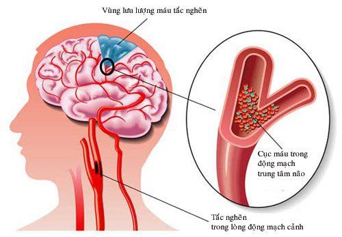
● This technique is indicated for patients with pathology of cerebral vascular malformations such as: cerebral aneurysm, cerebral arteriovenous catheterization, cerebral artery stenosis... or doctors need to evaluate blood supply vessels. intra- and extra-axial brain tumors.Experimental embolization to evaluate collateral circulation
Angiography is required for interventional radiology
Background erasing digital angiography system has no absolute contraindications. However, in cases where the patient has a blood clotting disorder, kidney failure, or a history of obvious allergy to iodinated contrast agents or pregnant women, the doctor will need to evaluate the condition more deeply before making a decision. can it be done?
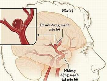
DSA được chỉ định đối với những bệnh nhân có bệnh lý về dị dạng mạch não như phình động mạch não
3. Process of digitizing brain angiography to erase background
The main performer will be a specialist doctor, adding an auxiliary doctor, radiology technician and nurse.An anesthesiologist and an anesthesiologist are needed to administer anesthesia and monitor the patient during the procedure.
3.1 Means to be used
● Digital erasure angiogram (DSA)● Specialized electric pump
● Film, film printer, image storage system
● Lead vest, apron, X-ray shielding.
3.2 Drugs used
● Local anestheticsPre-anesthesia and general anesthesia (if the patient has an indication for anesthesia)
Anticoagulants
Anticoagulant neutralizers
Water-soluble iodine contrast agents
Antiseptic solution for skin and mucous membranes
3.3 Common medical supplies
● Syringe 5; 10, and 20mlSyringe for electric pumps
● Distilled water or physiological saline
● Gloves, gown, cap, surgical mask
● Sterile intervention kit: Knife, scissors, tongs , 4 metal bowls, bean tray, tool tray
● Cotton, gauze, surgical tape
● Medicine box and first-aid kit for contrast contrast drug
● Special medical supplies
● Needle Arterial
Size 5-6F
Instrumentation Kit
Standard Lead 0.035'
Angiocardiography Catheter size 4-5F
Y Connector Kit
Three-prong Lock
Inlet Closure Kit
● The patient was met by a specialist and an anesthesiologist to explain the procedure carefully.
● You need to fast and drink 6 hours before performing digital brain angiography. You can drink a little water, but not more than 50ml of water.
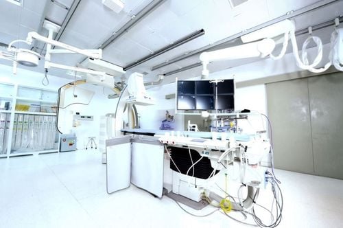
● In the intervention room: the patient lies on his back, fitted with measuring devices to control breathing, pulse, blood pressure, electrocardiogram, SpO2. Disinfect the skin, then cover with a sterile, perforated cloth.
4. Steps to conduct digital brain angiography erase background
4.1 Using the emotionless method
● Sterilize and anesthetize the puncture site● Needle puncture and catheterization
● For selective angiography of the internal carotid artery: Insert the catheter through the catheter into the internal carotid artery, inject contrast agent i- Iodine through the machine with a volume of 10ml, speed 4ml/s, pressure 500 PSI. The series of images and films focus on the skull in a straight, fully tilted, and 45 degree oblique position.
● For elective angiography of the external carotid artery: insert a catheter into the external carotid artery and inject iodinated contrast agent through the machine with a volume of 8ml, rate of 3ml/s, pressure of 500 PSI. Recording at 2-3 frames per second with a focus on the skull in a fully upright and tilted position.
● To select vertebral artery angiography: insert Vertebral 4-5F catheter, into vertebral artery (usually left side) inject contrast agent containing iodine, volume 8ml, speed 3ml/s, pressure 500PSI . The series of recordings and films focused on the posterior fossa skull in full tilt and upright position with the shadow at 25 degrees of cephalothorax, and with a 45 degree slant.
● 3D imaging can be performed depending on the pathology.
● After satisfactory scanning, withdraw the catheter and tube into the lumen and apply pressure directly on the needle puncture site for 15 minutes to stop bleeding. Then, apply pressure for 8 hours or can use a device to close the artery into the lumen.
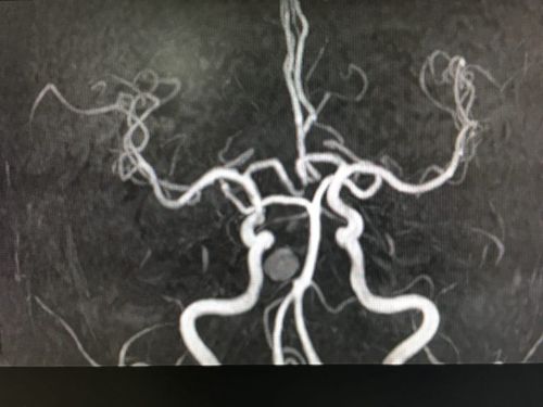
● Digital brain angiography image with background erasure will reveal the anatomical structures of the intracranial arterial system, including: internal carotid arteries, vertebral arteries, anterior cerebral arteries, middle brain and posterior brain on both sides. At the same time, the doctor will detect damage if any.
Currently, Vinmec International General Hospital also applies the technique of digital brain angiography to erase the background in the examination, diagnosis and detection of diseases for patients. All medical examination and examination procedures are performed by a team of doctors with many years of experience. Combined with that is a system of modern machinery and equipment. Thanks to good medical conditions, patients can be assured of the diagnosis results as well as the treatment direction when prescribed by the doctor.
Any questions that need to be answered by a specialist doctor as well as customers wishing to be examined and treated at Vinmec International General Hospital, you can contact Vinmec Health System nationwide or register online HERE.
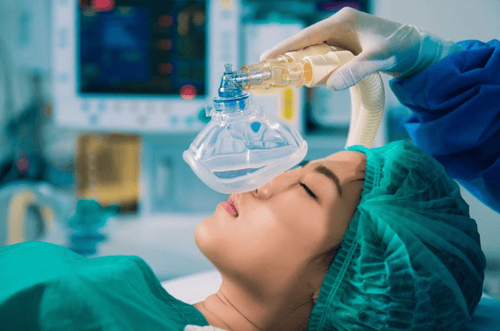
Trường hợp bệnh nhân quá kích động sợ hãi cần gây mê toàn thân khi làm thủ thuật
4.2 Carrying out digital brain angiography to erase the background
● Sterilize and anesthetize the puncture site● Needle puncture and catheterization
● For selective angiography of the internal carotid artery: Insert the catheter through the catheter into the internal carotid artery, inject contrast agent i- Iodine through the machine with a volume of 10ml, speed 4ml/s, pressure 500 PSI. The series of images and films focus on the skull in a straight, fully tilted, and 45 degree oblique position.
● For elective angiography of the external carotid artery: insert a catheter into the external carotid artery and inject iodinated contrast agent through the machine with a volume of 8ml, rate of 3ml/s, pressure of 500 PSI. Recording at 2-3 frames per second with a focus on the skull in a fully upright and tilted position.
● To select vertebral artery angiography: insert Vertebral 4-5F catheter, into vertebral artery (usually left side) inject contrast agent containing iodine, volume 8ml, speed 3ml/s, pressure 500PSI . The series of recordings and films focused on the posterior fossa skull in full tilt and upright position with the shadow at 25 degrees of cephalothorax, and with a 45 degree slant.
● 3D imaging can be performed depending on the pathology.
● After satisfactory scanning, withdraw the catheter and tube into the lumen and apply pressure directly on the needle puncture site for 15 minutes to stop bleeding. Then, apply pressure for 8 hours or can use a device to close the artery into the lumen.
● Digital brain angiography image with background erasure will reveal the anatomical structures of the intracranial arterial system, including: internal carotid arteries, vertebral arteries, anterior cerebral arteries, middle brain and posterior brain on both sides. At the same time, the doctor will detect damage if any.
Currently, Vinmec International General Hospital also applies the technique of digital brain angiography to erase the background in the examination, diagnosis and detection of diseases for patients.
Master. Dr. Le Hong Chien has many years of experience working in the field of diagnostic imaging and interventional radiology (endovascular intervention and extravascular intervention). Before being a Doctor of Diagnostic Imaging - Intervention at Vinmec Times City International Hospital, Dr. Chien was a Doctor at the Department of Diagnostic Imaging, Hospital 19.8 - Ministry of Public Security and Hong Ngoc General Hospital. .
Any questions that need to be answered by a specialist doctor as well as customers wishing to be examined and treated at Vinmec International General Hospital, you can contact Vinmec Health System nationwide or register online HERE.
SEE MORE
Digital background erasure angiography The principles of digital background angiography (DSA) Advantages of DSA background angiography