This is an automatically translated article.
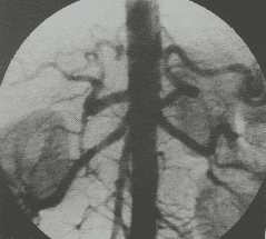
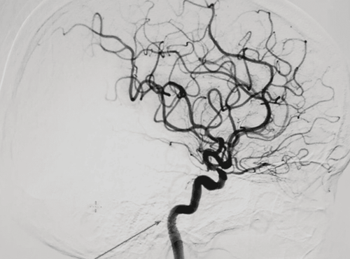
The article was professionally consulted by Specialist Doctor I Vo Cong Hien - Radiologist - Radiology Department - Vinmec Nha Trang International General Hospital.Cerebral angiography using a digitized background eraser allows the diagnosis and detection of cerebrovascular diseases or suspicion of accidental injuries causing brain artery damage.
1. What is a digitized scan of the cerebral arteries?
Cerebral angiography using a digital background eraser is an imaging technique that uses iodinated contrast to clear the background, thereby showing clearly the entire structure of the cerebrovascular system including arteries, veins and capillaries. circuit. Thereby, allowing detection and assessment of blood circulation in the brain.2. In what cases is a digital scan of the cerebral arteries indicated?
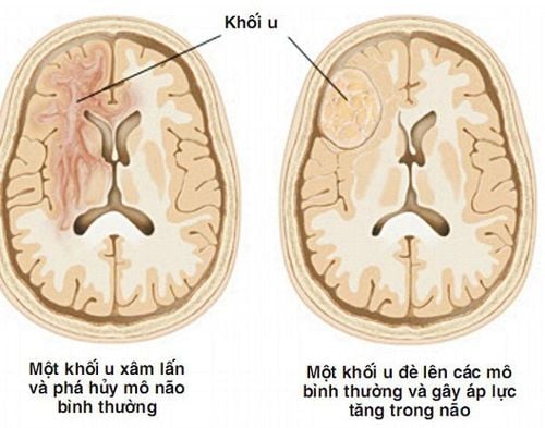
Cerebral angiography using a digital background eraser is indicated in the following cases:Diagnosis of diseases related to cerebrovascular malformations such as cerebral aneurysm, cerebral artery stenosis, cerebral arteriovenous catheterization, ...caused by vasculitis or atherosclerosis or acute cerebral artery occlusion, cavernous carotid artery dissection. Assess the blood supply to the brain tumor. Experimental evaluation of collateral circulation prior to embolization. Serving radiological intervention and diagnosis before interventional treatment.
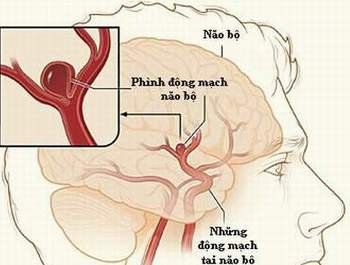
Bệnh nhân phình động mạch não được chỉ định thực hiện
3. How is digital scan of cerebral artery background done?
Digital scan of cerebral artery background is performed with the following steps:Step 1: Anesthetize the patient by placing the patient supine on the scanning table, setting up an intravenous line, and administering local anesthesia. For cases that cannot cooperate or are stimulated, fear can inject pre-anesthetic drugs for general anesthesia before the procedure. Step 2: The technique used to insert the catheter into the artery is Seldinger, the entry from the femoral artery. Only when access from the femoral artery is not possible should an alternative route of entry be selected, that is, the axillary artery, brachial artery, or the radial, primary carotid artery.

Bệnh lý động mạch não có thể được phát hiện bằng chụp số hóa xóa nền động mạch não
Cerebral angiography using a digital background eraser is a diagnostic imaging technique. Modern imaging allows doctors to diagnose diseases related to cerebral blood vessels and suspect accidental injury causing damage to cerebral blood vessels.
Digitization of cerebral artery background is an imaging technique that requires a high level of experience of the doctor and the perfect coordination of the patient. In order to achieve high diagnostic efficiency, patients need to choose reputable addresses that have digital scanning machines to erase the background and have modern and standard medical equipment from which to have timely treatment.
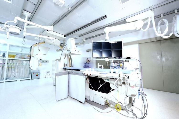
Vinmec có thực hiện kỹ thuật chụp số hóa xóa nền để chẩn đoán hình ảnh
BSCK I Vo Cong Hien has many years of experience in the field of imaging, especially in diagnostic ultrasound for general abdominal diseases, advanced cardiac - vascular, obstetrics and gynecology. Doctor Vo Cong Hien used to work at the Imaging Department of the University of Medicine and Pharmacy Hospital - Hoang Anh Gia Lai before working at the Imaging Department of Vinmec Nha Trang International General Hospital.
To register for examination and treatment at Vinmec International General Hospital, you can contact Vinmec Health System nationwide, or register online HERE.
In April & May 2021, when there is a need for examination and treatment of cerebral aneurysms at Vinmec Times City International Hospital and Vinmec Da Nang International General Hospital, customers will enjoy preferential treatment. Double:
- Free specialist examination and free x-ray
- 50% discount on costs for customers with appointment of post-examination treatment. The program is limited to the corresponding technique of each hospital and to customers who perform this treatment technique for the first time at Vinmec.
Please dial HOTLINE for more information or register for an appointment HERE. Download MyVinmec app to make appointments faster and to manage your bookings easily.


