This is an automatically translated article.
Digital subtraction angiography (DSA angiography) is a method performed for patients who are indicated for endovascular embolization intervention, endovascular aneurysm surgery, arterial stenting,... or in the diagnosis of vascular diseases.
1. What is background erasing digital angiography?
Digital subtraction angiography (also known as digital subtraction angiography) has the English name Digital subtraction angiography - DSA for short. This is a fluoroscopy endoscopic technique, used in interventional X-rays so that the doctor can clearly see the images of blood vessels. Radiation-blocking structures are digitally removed from the image, allowing blood vessels to be seen clearly.
A DSA angiogram system has the following structure:
X-ray generator; Image acquisition department; Image processing unit; Display department.
2. Principle of DSA
DSA is a combination of computerized image processing techniques and conventional angiography by Seldinger technique. The principle of digital angiography with background erasure is to use X-rays and fluorescent light to take angiograms at the necessary locations before and after the injection of contrast material into the blood vessels to be taken. . The computer then erases the background image, obtaining a full picture of the heart, carotid artery, abdomen, chest, and thighs - clarifying the vascular system.
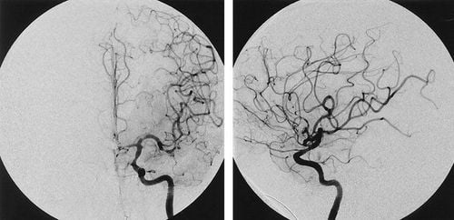
Chỉ định chụp DSA mạch máu não
Help doctors detect abnormal blood flow conditions early, thereby supporting accurate diagnosis and treatment of diseases related to blood circulation in the body. At the same time, the doctor can plan the surgery, pinpoint the exact location of the lesion in the body. As a result, many cases of cardiovascular disease, especially myocardial infarction, were identified, diagnosed and intervened promptly; The support of DSA angiography supports less invasive cardio-thoracic surgery. Patients will reduce pain, reduce bleeding, shorten the time of mechanical ventilation, recuperation, shorten hospital stay and ensure aesthetics compared to open surgery.
3. Indication to perform background erasure digitized angiography
Digital background erasure is indicated in cases where interventional radiology is effective for vascular procedures. Specifically, a DSA is performed for patients who:
Balloon angioplasty; Trans-arterial aneurysm surgery; Placement of arterial stents; Intravascular embolism; Remove blood clots. In addition, digital angiography erasing the background is also used in the diagnosis of vascular diseases such as:
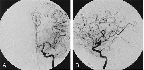
Brain aneurysm, especially intracranial aneurysm; Blockage in the lumen of a vein or artery; Blood vessel bleeding; Arteriovenous malformations ; Vascular study of melanoma.

Chụp số hóa xóa nền được áp dụng để chẩn đoán các bệnh mạch máu
4. Process of performing background digitizing angiography
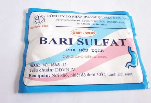
4.1 Before performing The patient fasts for breakfast; Carry out necessary tests including: blood test, electrocardiogram measurement, chest X-ray; The doctor explains to the patient about the procedure of performing a DSA scan, the risk of possible complications; The patient lies in a chair, the X-ray device moves around. 4.2 During the procedure, the patient is subjected to routine X-ray, obtaining images of normal anatomical structures and radiation-blocking objects such as surgical pins, stents,... Images obtained from this step is used as background image; The patient is injected with a special dye to highlight blood vessels in the legs, heart, or other organs. Contrast will stand out on X-ray films when hit by high-density X-rays; X-rays are beamed through the body and are picked up by a bulb. A lens is placed between the bulb and the video camera to limit the intensity of light reaching the camera; As the contrast material enters the patient's body, the machine acquires the animation in a preset time unit. Then, the image processing unit will take the received image, excluding the background image.

Tiêm chất cản quang để làm nổi bật mạch máu khi chụp
4.3 After performing the DSA angiography patient is given a catheter into the lumen from the groin to the artery to be angiographically in order to inject the contrast agent. Therefore, digitized angiography to remove the background is an invasive imaging technique with a high risk of complications. Therefore, patients need to follow the doctor's instructions during the DSA scan. After the scan, the patient was bandaged in the groin area, lying motionless for 24 hours and absolutely avoiding leg movement.
From the results obtained, the doctor will have more bases to make a diagnosis or prescribe treatment for the health problems that the patient is experiencing.
4.4 Risk of complications
The DSA scan is usually painless. Some complications may occur after digital angiography erasing the background, including:
Complications at the injection site: Blood clot formation, pseudoaneurysm, arteriovenous fistula, damage to adjacent tissues; Systemic complications: thromboembolism, nephrotoxicity, air embolism, vascular dissection. Background digitized angiography is an important technique in the diagnosis and treatment of many vascular diseases. When performing the technique, the patient should follow all instructions of the doctor to achieve high therapeutic effect and reduce the risk of complications.
Vinmec International General Hospital with a system of modern facilities, medical equipment and a team of experts and doctors with many years of experience in medical examination and treatment, patients can rest assured to visit. examination and treatment at the Hospital.
To register for examination and treatment at Vinmec International General Hospital, you can contact Vinmec Health System nationwide, or register online HERE.