Overview of acromioclavicular joint dislocation
They can only partially slide across each other because of the clavicle joint, which is a flat moving joint. A network of ligaments and capsular ligaments surround the joint. The sacroclavicular ligament system, the clavicle ligament (conical ligament, ladder ligament), and the surrounding deltoid and trapezius muscle system are the components that anchor the clavicle joint.
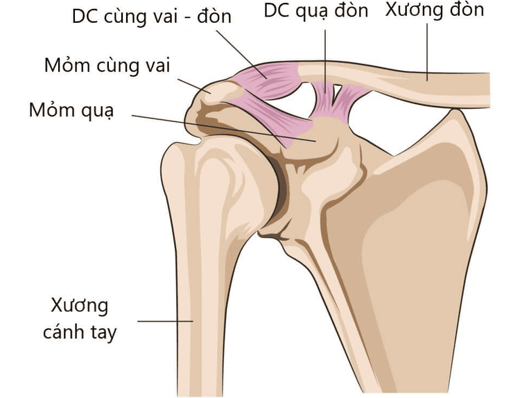
Acute sacroclavicular dislocation is a condition that develops following trauma (within three weeks), destroys the ligamentous system that stabilizes the joint, and causes the lateral clavicle to dislocate above its normal position. There are two different injury mechanisms that can result in clavicle dislocation: the direct mechanism, in which the patient falls and strikes their closed shoulder against the hard ground, pushing their sacroiliac process inward and downward; and the indirect mechanism, in which the patient falls against the hard ground, the force is transferred to the joint, and the shoulder blade injures them.
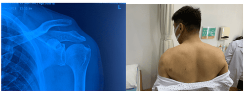
Shoulder dislocation has been identified
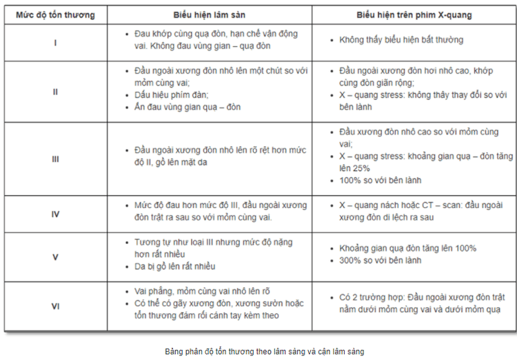
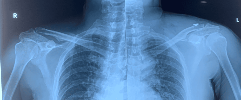
Clinical pain, palpation of the lateral clavicle sticking out above the skin, the Piano key (+) sign, and radiographs that compare the bilateral shoulder apex are all used to make the diagnosis of sacral clavicular dislocation. It will be more challenging to diagnose grade I and II lesions clinically and on X-ray films, and additional computed tomography or magnetic resonance imaging may be needed.
Grade of the shoulder blade's dislocation?
Treatment of dislocation of the shoulder blade
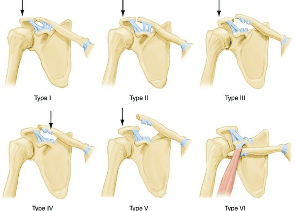
Lesions of type I and type II will require conservative treatment, whereas lesions of type III and above will require surgical intervention.
There are several surgical procedures for treating clavicle dislocation, including fixed needle piercing, the Bosworth technique, and the use of hook plate splints. These procedures have comparatively good outcomes but also come with numerous risks and inconveniences for the patients.
There are currently various surgical methods available to totally endoscopically repair the clavicle joint thanks to the development of shoulder arthroscopy.
We are performing endoscopic fixation of the acromioclavicular joint at the upper extremity surgery section of the Vimec Hospital Orthopedic Trauma Center utilizing sutures and tightropes.
Arthroscopy technique of shoulder joint surgery
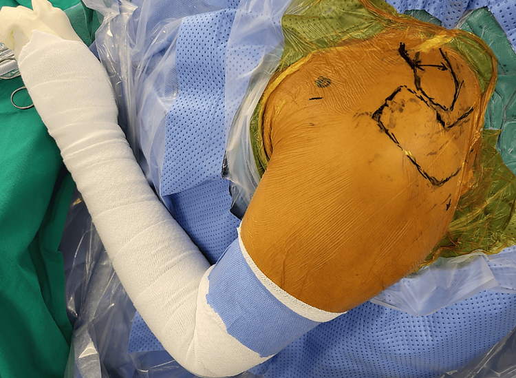
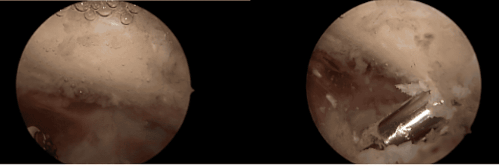
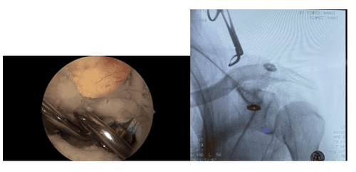
Anesthesia was administered to the patient, who was then positioned in a beach chair. Two ports were used to enter the shoulder joint and check for injury in other shoulder joints. The crow's jaw is then readily seen. Using a finder, cut a tunnel through the clavicle and crow's feet, insert tightrope and thread, and fasten the joint with the clavicle. To assess the outcomes, conduct a test immediately on the C-arm.
Advantages of laparoscopic surgery for treating shoulder joint dislocation
The laparoscopic incision is smaller and more aesthetically pleasing; the patient experiences less discomfort and returns to normal life more quickly; yet, the complication rate is relatively low.
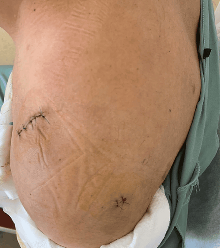
Vinmec Hospital's proficiency in shoulder arthroscopy treatment of shoulder blade dislocation.
With many years of combined expertise in shoulder arthroscopic surgery in general and shoulder dislocation surgery specifically, our team consists of top experts. A group of specialists in rehabilitation also creates workouts every day and for patients after they are discharged.
Equipment and machinery for shoulder joints that are completely specialized. The endoscopic procedure's use of cold water helps patients control bleeding and recover from surgery quickly.
Please dial HOTLINE for more information or register for an appointment HERE. Download MyVinmec app to make appointments faster and to manage your bookings easily.


