This is an automatically translated article.
The article is professionally consulted by Master, Doctor Ton Nu Tra My - Department of Diagnostic Imaging - Vinmec Central Park International General Hospital.Breast ultrasound is a safe, effective, low-cost technique that can be performed in all subjects. Breast ultrasound helps to detect abnormal breast features early for timely treatment.
1. Breast ultrasound
Breast ultrasound is a technique that uses ultrasound waves to create images of the internal structure of the breast. This is a safe, effective, low-cost, painless, non-toxic method that can be used by everyone from the elderly to children. Due to its many advantages, breast ultrasound is currently a very commonly used method, helping to detect breast abnormalities early.Breast ultrasound should be done periodically once a year, or as soon as the body has abnormal symptoms such as:
Abnormally large breasts, chest pain There are lumps in the breast, nipples are retracted in the Area skin around the nipple changes appearance of lymph nodes under the armpit
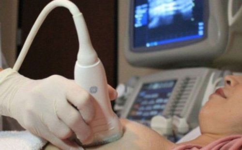
Siêu âm vú nên được thực hiện định kỳ mỗi năm 1 lần
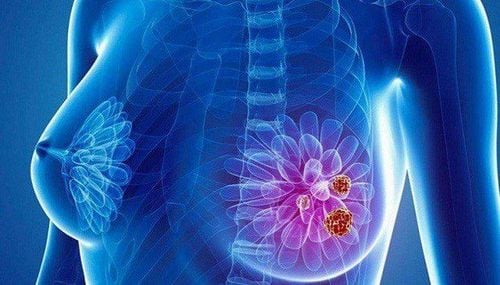
2. Abnormal breast features detectable by ultrasound
Breast tissue is made up of many components such as connective tissue, fatty tissue, milk glands, milk ducts. In young non-breastfeeding women, the normal breast parenchyma structure consists of fibrous parenchyma, with little or no subcutaneous fat. As age and number of births increase, more and more fat is deposited in the subcutaneous fat layer and the fat behind the breast. This is a normal phenomenon of the body.When doing breast ultrasound, the doctor can detect some abnormal features of the breast such as:
2.1. Breast cysts Breast cysts are fluid-filled sacs in the breast that are the most common cause of palpable breast masses in women aged 35-50 years. Women can have one or more breast cysts, in one or both breasts. The causes of breast cysts are tubular lobular obstruction, fibrosis, or intraductal epithelial proliferation.
The mammary cyst seen on ultrasound is a well-defined, oval or round, thin-walled structure that allows sound waves to pass through. Breast cysts can be simple cysts or complex cysts. A mammary cyst is considered complex when the doctor sees a faint echo or internal debris. These blunt echoes can be calcium crystals, cholesterol crystals, pus, blood,...
2.2. Mammary duct ectasia Mammary duct ectasia is a disease involving the main milk ducts of the mammary glands. It usually occurs in women aged 40-50 who have had many children and have breastfed for many years.
Under breast ultrasound imaging, dilated milk ducts may present as a single tubular structure filled with fluid or with condensed debris. Tubular debris may present as focal lesions. If debris fills the lumen, it can be mistaken for a solid mass.
2.3. Fibroadenoma is a benign tumor in the breast.
Breast fibroids are common in women under 35 years of age, caused by a disturbance in the balance of estrogen and progesterone hormones. Tumor size is usually less than 5cm, can have many tumors and appear in both breasts.
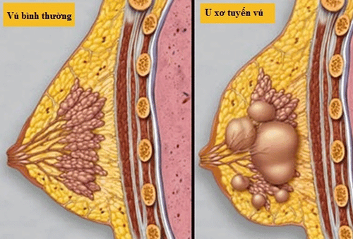
U xơ tuyến vú là một dạng khối u ở vú lành tính xuất hiện khi có một số tế bào tăng sin
2.4. Chronic abscess of the breast Chronic abscess of the breast is usually located centrally or below the areola. Patients feel pain, fever, fatigue, blood test will have diphtheria increased. Under breast ultrasound imaging, abscesses often present as well-defined or poorly defined structures, containing echogenic fluid, posterior echogenicity with echogenic inflammatory infiltrates, and diffuse hyperperfusion of the breast tissue. around.
2.5. Fibrocystic changes: Fibrocystic changes or cystic fibrosis of the breast can have many different characteristics depending on the stage and degree of morphological changes. In the early stages of the disease, ultrasound of the breast may be normal, although a palpable breast lump may be seen on physical examination. In subsequent stages, focal areas of parenchymal thickening may be seen, which may be accompanied by single cysts or clusters of small cysts.
2.6. Lipoma A lipoma is a soft breast lump that can deform with pressure on the transducer and grows slowly. Lipoma is a well-defined tumor, under the ultrasound image, the tumor has a capsule, echogenic structure similar to adipose tissue.
2.7. Phyllodes tumor is a slow-growing breast tumor, mostly benign, although usually large in size, can occupy the entire mammary gland. Ultrasound imaging often shows well-defined mass, heterogeneous echogenicity, and sometimes degenerative internal cysts.

Áp xe mạn tính ở vú thường nằm ở vùng trung tâm hoặc dưới quầng vú
Breast cancer screening package at Vinmec for the following subjects:
Female customers, over 40 years old. Customers wishing to be able to screen for breast cancer Customers are at high risk of cancer – especially customers with a family history of breast cancer. Women of reproductive age, perimenopause and menopause. Women who are having symptoms of breast cancer such as: pain in the breast, lump in the breast, etc
Please dial HOTLINE for more information or register for an appointment HERE. Download MyVinmec app to make appointments faster and to manage your bookings easily.
SEE MORE
Everything you need to know about breast biopsies Why should breast biopsies be guided by ultrasound? It takes time for the immune system to recover from breast cancer chemotherapy