This is an automatically translated article.
An ultrasound-guided breast biopsy is performed when an abnormal area in the breast is so small that it is sometimes not obvious by hand palpation. Ultrasound technology helps support breast biopsies to detect cancer or other breast diseases early.1. What is an ultrasound-guided breast biopsy?
A breast biopsy is done to remove some cells from a suspicious area in the breast and examine them under a microscope to arrive at a diagnosis of whether the cells are benign or malignant. An ultrasound-guided breast biopsy is performed when an abnormal area in the breast is so small that it is sometimes not obvious by hand palpation.The doctor will conduct the biopsy under the guidance of the ultrasound machine by holding the ultrasound probe in one hand and the needle in the other hand. The ultrasound machine is responsible for diagnosing the route of the needle to the abnormal area in the breast tissue, from which to determine the location of the biopsy needle. This helps the biopsy needle to reach the right place to take cells and not leave scars or affect the patient's aesthetics.
2. When is an ultrasound-guided breast biopsy done?

Bác sĩ chỉ định làm sinh thiết vú khi khám lâm sàng, siêu âm vú, chụp X-quang vú phát hiện ra khối u vú đặc nghi ngờ
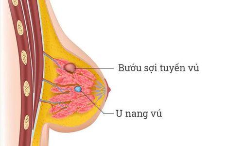
Suspicious solid breast tumor Structural disorder of breast tissue A variable area Abnormal changes of glandular tissue, suspected breast cancer. Ultrasound-guided breast biopsy technique is indicated in the following cases:
Lesions with BIRADS classification 4 and 5 Axillary nodes with suspected metastasis Axillary nodes, recurrent lesions Lesions cystic form suspected of malignancy; Lesions require aspiration before biopsy. Under the guidance of ultrasound, the doctor can perform three procedures:
Fine-needle aspiration, which uses a very fine needle to aspirate cells in the abnormal area Core biopsy uses a large-aperture needle to remove a sample tissue after each puncture Localize with a hook wire, place a needle in the suspicious area to help the surgeon locate the lesion when performing the biopsy procedure.
3. The advantage of the trick brings
Guide to ultrasound to evaluate, diagnose, differentiate fluid-filled cysts from solid tumors Helps detect very small lesions that cannot be felt or seen with the naked eye Ultrasound is a safe diagnostic tool for in pregnant women for whom other means are not available Identify solid breast lumps in patients about 35 years of age, in cases where mammography results are unclear. In addition, breast ultrasound also helps to timely detect the growth of cancer inside, causing thickening of the skin in the breast. In these cases, it is usually indicated to perform a breast biopsy under ultrasound guidance.Breast ultrasound helps guide the needle for biopsy, helping the patient feel more comfortable, not having to put too much pressure on the breast compared to mammography.
4. How does the patient feel during and after the biopsy?

Tại vùng sinh thiết, vài ngày sau đó, bệnh nhân có thể bị sưng và thâm tím do chảy máu dưới da
After performing the biopsy If there is pain, the patient can use ordinary pain relievers. At the biopsy site, several days later, the patient may experience swelling and bruising due to bleeding under the skin. Patients should not be concerned with this phenomenon. If there is swelling, bleeding, discharge, redness, or a burning sensation after the biopsy, the patient needs to see a doctor immediately.
Currently, Vinmec International General Hospital is one of the very few hospitals in the country that applies the technique of breast biopsy with vacuum-assisted vacuum aspiration under Stereotactic positioning. This technique is more effective than the core needle biopsy method: Thanks to the use of a biopsy needle with a large size of 12 G or more, combined with vacuum suction, it helps to get more samples, thereby making it possible to obtain more samples. higher probability of success. In addition, this technique is also less invasive than the open biopsy method: The incision site does not require any recovery stitches, does not require special care, and the patient can return to normal daily life right away. tomorrow. In addition, the placement of markers to mark the intervention site is carried out right in the procedure to help manage the lesions after the test results are more favorable.
Please dial HOTLINE for more information or register for an appointment HERE. Download MyVinmec app to make appointments faster and to manage your bookings easily.


