This is an automatically translated article.
The article is professionally consulted by MSc, BS. Dang Manh Cuong - Doctor of Radiology - Department of Diagnostic Imaging - Vinmec Central Park International General Hospital. The doctor has over 18 years of experience in the field of ultrasound - diagnostic imaging.Digital erasure of the head and neck angiogram is an imaging technique with iodinated contrast injection through the catheter to show dynamic and static images of the head, face and neck.
1. When is digital cerebral angiography indicated?
Angiography of the head, face, neck and neck with digital erasure of background injecting iodine-containing contrast agent will be prescribed by the doctor when the patient has vascular malformations in the head and neck area: Arteriovenous catheterization, pseudoaneurysm ...The doctor needs to evaluate the tumor blood vessels in the head, face, and neck of the patient. The patient has bleeding due to impact causes such as trauma, tumor invasion, nose bleeding Need to evaluate the collateral circulation Need angiography for interventional radiology. Digital angiography of the head, face and neck, erasing the background of iodine-containing contrast injection, has no absolute contraindications. However, in some cases with coagulation disorders, renal failure, a history of obvious allergy to iodinated contrast agents, pregnant women need a closer evaluation of the doctor for a more thorough evaluation. before giving this indication.

Chụp mạch vùng đầu mặt cổ số hoá xoá nền tiêm thuốc đối quang chứa i ốt sẽ được bác sĩ chỉ định khi bệnh nhân mắc các bệnh lý dị dạng mạch vùng đầu cổ
2. Digital angiography of head, face, neck, erasing background injecting iodine-containing contrast agent
2.1 Preparation
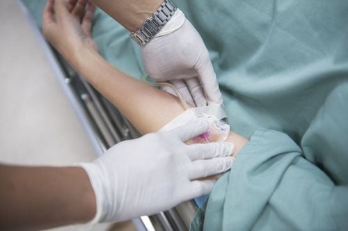
The patient was thoroughly explained by the doctor about the procedure to coordinate with the doctor. Need to fast, drink before 4-6 hours before the procedure. Can drink no more than 50ml of water In the intervention room: The patient lies on his back, has a machine to monitor breathing, pulse, blood pressure, electrocardiogram, SpO2 Disinfect the skin, then cover with a sterile cloth with holes. including: 1 specialist, 1 assistant doctor, anesthesiologist (if the patient has an indication for anesthesia), radiology technician, nurse Selecting an anesthetic method Place the patient in supine position scan table, intravenous line (usually 0.9 % isotonic saline serum), pre-anesthetic injection, with the exception of young children (under 5 years old) who have not had a sense of cooperation or are too scared General anesthesia is required for the procedure. Select a technique using the Seldinger technique. The entrance of the catheter into the lumen can be from the femoral artery, axillary artery, brachial artery, the main carotid artery, and the radial artery. Usually most are from the femoral artery, unless this is not possible, other access routes are used.
Kỹ thuật chụp mạch số hoá nền DSA
2.2 Technical implementation
Disinfection and local anesthesia Needle puncture and arterial access For selective external carotid angiography: Insert an arterial catheter to the external carotid artery and inject contrast agent through the machine at a volume of 8 ml, at a rate of 3 ml/s , pressure 500 PSI. Recording and taking series of films (series) focusing on the skull in a fully upright and tilted position. For selective angiography of the internal carotid artery: Insert an arterial catheter through the lumen into the internal carotid artery and inject contrast medium through the machine with a volume of 10 ml, rate of 4 ml/s, pressure of 500 PSI. Imaging and radiographs of the cranial focus series upright, fully tilted, and 45 degrees oblique For selective vertebral artery angiography: insert a Vertebral 4-5F catheter, to the vertebral artery (usually lateral) left) contrast injection, volume 8ml, speed 3ml/s, pressure 500PSI. Mass imaging and imaging (series) focusing on posterior fossa craniofacial position in full tilt and upright position with shadow 25-degree oblique, and 45-degree eccentricity. 3D angiography can be performed depending on the pathology For subclavian angiography: Insert the catheter into the subclavian artery or selectively into the biceps and scapula to inject drugs. After satisfactory scanning, remove the catheter, remove the vascular catheter (catheter) from the lumen, apply pressure directly on the needle puncture site for about 15 minutes to stop bleeding, then apply pressure for 6 minutes. hour. Angiography of the head and neck area digitalized erasing the background injected with iodinated contrast agent, the image results clearly show the anatomical structures of the internal carotid artery, external carotid artery, vertebral artery, cerebral artery system anterior, midbrain and posterior brain on both sides. Detect damage, if any.Please dial HOTLINE for more information or register for an appointment HERE. Download MyVinmec app to make appointments faster and to manage your bookings easily.
SEE MOREDigital imaging to erase the background of the brain's arteries Digitally remove the background and treat brain aneurysms with flow change Digital scan to erase the background and dural arteriovenous catheterization