This is an automatically translated article.
The article is professionally consulted by Master, Doctor Vu Thi Hau - Radiologist - Department of Diagnostic Imaging and Nuclear Medicine - Vinmec Times City International General Hospital.Computed tomography is a simple, non-invasive technique with high diagnostic efficiency. When taking, depending on the case, the use of contrast agents may be indicated. Contrast is used in computed tomography to help clinicians see the lesion clearly, thereby providing appropriate treatment methods for each case.
1. Maxillofacial computed tomography with contrast injection in the axial and coronal planes
Computed tomography of the face with contrast injection in the axial and coronal planes is a technique to introduce a volume of contrast agent into the body through an intravenous line. Then take pictures with the machine to examine the diseases of the maxillofacial region, the nose and sinuses, the throat... To determine the diagnosis for conventional X-ray techniques in two planes, the horizontal plane (axial) and vertical (coronal).For maxillofacial computed tomography using contrast, there are two directions: axial (transverse section) and coronal (horizontal) plane:
Axial direction: This is the cutting direction from the upper border. frontal sinus to the end of the mandibular arch. Equal to the hard palate. The coronal direction cuts from the frontal sinus to the posterior border of the mandible, perpendicular to the transverse direction. When taking pictures, it is necessary to take pictures in these two directions to investigate the lesion thoroughly.
For modern multi-sequence CT scanners, axial thin tomography and coronal reconstruction bring many advantages such as faster capture time, reduced X-ray dose, better image quality,...
2. Indications and contraindications
Indications:Injuries to the maxillofacial region.Inflammation, infection, abscess Tumor lesions in the maxillofacial region According to the professional requirements of the treating doctor. Contraindications:
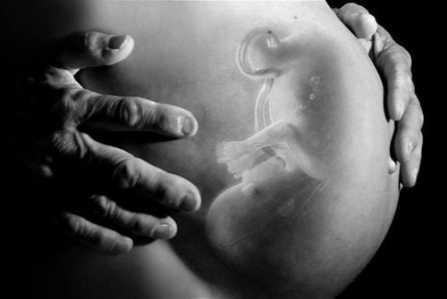
Relative contraindications: Pregnant women, especially in the first 3 months, young children. Pregnant women must use a lead shirt to cover the abdomen if taking pictures. Patients with kidney failure, liver failure, severe heart failure, chronic diseases such as diabetes, bronchial asthma; have a history of allergy to contrast media...

Phương pháp này chống chỉ định với bệnh nhân suy thận
3. Procedure for facial computed tomography
3.1 Preparation
Performer: To take a computed tomography scan of the maxillofacial region, it is necessary to have a doctor and a technician specializing in imaging.Means used to take computed tomography of the maxillofacial region include:
Computerized tomography machine, electric injection pump. Film, printer and image storage system. Water-soluble iodinated contrast agent. Medical supplies: Syringes, needles, syringes for electric injection pumps, antiseptic solution, physiological saline, gloves, masks, tray sets, cotton swabs. Medicine box and tools to manage complications in case of abnormality when the patient injects contrast agent. Patient:
The patient is clearly explained about how to take computed tomography, notes when taking the scan and possible complications so that he can coordinate with the person taking the scan. Remove items that can cause image interference such as earrings, necklaces, hairpins... You need to fast for 4 hours before taking pictures. You can drink but not more than 50ml of water. Patients who are overstimulated, cannot lie still, are anxious and afraid, or in the case of young children who can't move and are uncoordinated: Need to give sedation before the scan.
3.2 Steps to take
Computed tomography scan in both axial and coronal directions. For facilities with multi-slice computerized tomography machines (from 16 rows or more) it is possible to perform only the axial (axial) section, and then reconstruct the horizontal and other directions, but still ensure ensure the same image quality as the cross-sectional image quality.Perform imaging in two directions before contrast injection:
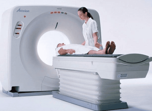
Máy chụp cắt lớp vi tính
3.2.1 Transverse (axial) direction
Patient position: Lying on the back, with the head in the middle, the patient needs to lie still, not moving because it will cause image noise. Perform positional shooting, shooting in a plane parallel to the hard palate. Taken horizontally, layer by layer from the base of the skull to the hyoid bone. The thickness of each cut layer is from 3mm-5mm. The jump is equal to the layer thickness, spiral cutting is recommended.3.2.2 Horizontal (coronal) orientation
Patient position: The patient lies supine with the head supine maximally or supine maximally supine. The patient needs to lie still for a few minutes while the technician takes the scan. Perform positioning capture, according to the cutting plane perpendicular to the cross-sectional plane. Photograph in the vertical direction of each section from the tip of the nose to the posterior spine of the cervical spine. The thickness of each cutting layer is 3mm-5mm. The jump is equal to the layer thickness, spiral cutting is recommended. Perform cross-sectional imaging after contrast injection with the same procedure and imaging method as without contrast injection.After taking: Determine the standard image and print the results in both cross-sectional and horizontal directions before, after contrast injection, in both bone and software windows.
3.3 Evaluation of results
The doctor reads the results, describes the lesion: Location, structure, size, spread of the lesion... Compare computed tomography images and clinical signs Provide diagnostic orientations. Recommend other examinations to coordinate. The doctor can provide additional professional advice to the patient upon request.4. Accidents and how to handle accidents when shooting
Young children may not cooperate during the imaging process: Treat by taking pictures while the child is asleep, using sedation or anesthesia depending on the case. The patient is unable to tilt the neck for the coronal scan, can reconstruct the image from the cross-sectional direction with multi-slice machines, in which case the thinnest helical transverse section is recommended. possible, to reproduce the best image. For the method of computed tomography of the maxillofacial region, contrast is used, so there may be complications caused by contrast. Therefore, before taking the patient, it is necessary to declare a complete medical history to limit the risk of complications.Master. Doctor. Vu Thi Hau has many years of experience in the field of diagnostic imaging including magnetic resonance, CT and ultrasound. Graduated from Hanoi Medical University in 2014 with a BSNT and trained in France for 1 year. Currently engaged in the field of diagnosis of neurological, head and neck, ear - bone, musculoskeletal, digestive and urological diseases.
Any questions that need to be answered by a specialist doctor as well as customers wishing to be examined and treated at Vinmec International General Hospital, you can contact Vinmec Health System nationwide or register online HERE.
SEE MORE
Maxillofacial computed tomography: What you need to know Cerebral angiography for early diagnosis of brain stroke What are the benefits of Magnetic Resonance Imaging (MRI)?


