This is an automatically translated article.
The article is expertly consulted by Master, Doctor Luu Thi Bich Ngoc - Doctor of Radiology - Department of Diagnostic Imaging and Nuclear Medicine - Vinmec Times City International General Hospital.Subframe computed tomography with contrast injection is a technique that uses an X-ray computerized tomography machine to capture the subframe. Computerized tomography subframe allows assessment and detection of pathological abnormalities of organs such as ovaries, uterus, bladder, prostate, ...
1. What you need to know about contrast-enhanced computerized tomography
Subframe computerized tomography with contrast injection is a technique that uses X-rays of a low-receiver system to examine the pelvic region. The results after capturing and processing, reconstructing images allow structural assessment and detection of abnormalities and pathologies of the pelvic organs such as: ovaries, uterus, bladder, prostate, etc. ..2. When is a subframe computed tomography with contrast injection indicated?
Computed tomography of the subframe with contrast injection is indicated in the following cases:● Gynecological diseases such as: Gynecological inflammation, gynecological abscess.
● Unexplained abdominal pain in the pelvic region such as appendicitis, diverticulitis, abscess inside the pelvis.
● Tumors in the ovary, uterus, bladder, prostate, subperitoneal cavity.
● Other abnormalities such as: Vaginal - bladder fistula, vagina - rectum, ...
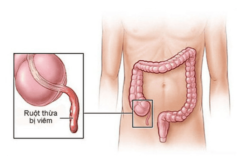
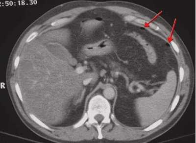
Bệnh nhân viêm ruột thừa được chỉ định chụp cắt lớp vi tính tiểu khung có tiêm thuốc cản quang
3. Steps to conduct subframe computed tomography with contrast injection
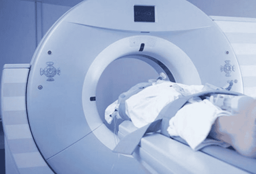
The procedure of subframe computed tomography with contrast injection includes the following steps:● Step 1: The patient is lying supine on the table, raising both arms above the head to avoid image interference. Instruct and ask the patient to hold their breath during the scan to avoid image noise.
● Step 2: Before injecting contrast, take cross-sectional layers of the subframe to locate the lesion. Measure the density of the suspected lesion for the purpose of evaluating the composition of the lesion with fat, calcification, and bleeding. The results are used to compare with the density of the lesion area after contrast injection to help assess the drug permeability of the lesion area more or less. The cut layers taken before and after injection have a thickness of 5-8mm, with small lesions the cut thickness should be 3mm.
● Step 3: Inject the contrast agent with a dose of 1.5 - 2 ml/kg of body weight, use an injection pump to inject quickly with a minimum injection speed of 3ml/s, in addition, an injection pump It also helps to accurately control the arterial and venous rhythms after injection. Depending on the size of the individual change the field of view (FOV) accordingly. The width of the window is varied and adjusted so that the entire skeletal system, fat, air and soft tissue can be assessed.
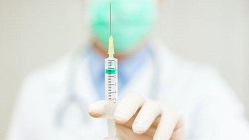
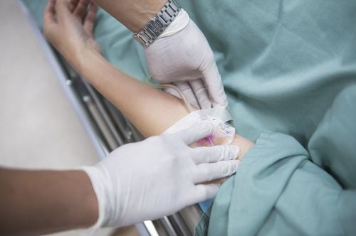
Tiêm thuốc cản quang với liều lượng là 1,5 - 2 ml/kg cân nặng khi tiến hành chụp cắt lớp vi tính
● Step 5: Computed tomography of the subframe of the layers in the venous phase pulse after injection from 60 - 70 seconds to assess drug clearance of tumor lesions, detect lesions of the rupture or contusion in visceral trauma, assess drug absorption of the tube wall digestion as well as the wall of an abscess.
● Step 6: Depending on the case, the subframe computed tomography is 5-7 minutes later after injection. In some cases of lower third ureteral calculi, or suspected low urinary tract fistula, imaging may be performed very late, when the bladder is distended to expel urine containing iodinated contrast.
● Step 7: Recreate the image on the camera at the shooting chamber. The image results must be clear and free of noise to serve the diagnosis.
Master, Doctor Luu Thi Bich Ngoc has 06 years of experience working in the field of diagnostic imaging, received formal training and graduated with a Master's degree from Hanoi Medical University. Dr. Ngoc has strengths in diagnosing breast, thyroid, liver cancers early. experience in doing ultrasound of tissue elastography for liver, breast, thyroid diseases... From 2019 until now, Dr. Ngoc has worked at Vinmec Times City International Hospital with a position at the department. Image analysation.
Any questions that need to be answered by a specialist doctor as well as customers wishing to examine and perform a subframe computed tomography with contrast injection at Vinmec International General Hospital, you can contact us. Vinmec Health System nationwide or register online HERE.
SEE MORE
Pelvic magnetic resonance imaging (pelvic region): What you need to know Factors causing pelvic pain, inflammation Does magnetic resonance imaging (MRI) have any effect?