This is an automatically translated article.
The article was written by doctors at the Radiation Oncology Center - Vinmec Times City International General HospitalComparison of repositioning of patients undergoing radiation therapy for pelvic cancer on two immobilizers with Vac-lock and without Vac-lock, using Cone - Beam CT at Vinmec Times International General Hospital City. Understanding the process will help customers have a more realistic and objective view.
1. Summary of patient repositioning of two immobilizers with Vac-lock and without Vac-lock
Purpose of comparing the patient position reconstruction of two immobilizers using Vac-lock and not using Vac-lock in radiotherapy for pelvic tumors at Vinmec Times City Hospital.
Subjects and methods: A total of 26 patients with VMAT radiotherapy for pelvic cancer were selected from January 2018 to September 2020 at Vinmec Times City International Hospital, the patients were divided into two groups: Group I included 13 patients receiving VMAT radiotherapy using Vac-lock. Group II included 13 patients receiving VMAT radiotherapy without Vac-lock. Patients received 2D-kV verification scans daily or twice a week for 3D-CBCT images. A total of 633 image acquisitions verified the repositioning of the two patient groups. Pairs of 2D-kV and 3D-CBCT images were analyzed and re-positioned in three dimensions anterior-posterior (AP: anterior-positior) and left-right (LR: left-right) and top-infrior (superior-infrior). ), error data are stored on the ARIA system (Varian Medical System, CA, USA). These data were used to evaluate setting errors in the LR, SI, and AP directions, and to compare patient position reconstruction in two different fixation kits.
The results of the average setting error re-establishing the treatment position of all patients in the AP, SI, and LR directions, respectively, are: (1.45 ± 2.05mm); (1.58 ± 3.22mm); (1.55 ± 1.82mm). The average setting error re-positioning the correct position of group I using Vac-lock in the AP, SI, LR directions is: (1.30 ± 2.12mm), respectively; (1.19 ± 3.29mm); (1.54 ± 1.67mm). The average setting error of correct position reproducibility of group II without Vac-lock in AP, SI, LR directions is: (1.39 ± 1.99mm), respectively; (1.87 ± 3.16mm); (1.54 ± 1.98mm).
Conclusion accurate repositioning of the treatment position when using immobilizer kit with Vacklock gives better repositioning result than without using Vac-lock. Priority is given to the use of a Vac-locked subframe immobilizer to stabilize the patient's position and accurately re-establish the daily treatment position in all AP axes; SI and LR in daily clinical radiotherapy practice.
Keywords: Installation error, radiotherapy for cancer of the pelvis, patient immobilizer, OBI system ...
COMPARISON OF REPRODUCIBITY OF PATIENTS BY USING THE TWO IMMOBILIZATION DEVICES WITH VAC-LOCK AND NON-VAC patients-LOCK FOR PELVIS RADIOTHERAPY BY THE CBCT SYSTEM AT VINMEC TIMES CITY INTERNATIONAL HOSPITAL
ABSTRACT
Purpose: To compare the purpose of two accuracy sets of the immobilization devices for pelvis radiation therapy at vimec international hospital. Materials and Methods : A total of 26 patients with VMAT technical radiotherapy with pelvis cancers were selected from January 2018 to September 2020 at Vinmec Times City hospital, patients were divided into two groups: Group I includes 13 patients Technical radiation therapy VMAT using Vac-lock ; Group II includes 13 patients with radiotherapy technology VMAT not using Vac-lock. Patients were scanned for verification 2D-kV daily or twice a week for 3D-CBCT images. A total of 633 imaging attempts verified the repositioning of the two groups of patients. 2D-kV and 3D-CBCT image pairs analyzed and evaluated for repositioning anterior-posterior (AP: anterior-posterior) and left-right (LR: left-right) and superior-inferior (superior-inferior) ), setup error data is stored on ARIA system (Varian Medical System, CA, USA). These data were used to evaluate the setting error in the LR, SI, AP directions, and compare patient repositioning in two different immobilization devices system. Results: The average setup error of reproducibility treatment position of all patients in AP, SI, LR directions respectively was: (1.45 ± 2.05mm); (1.58 ± 3.22mm); (1.55 ± 1.82mm). Accurate repositioning average setup error of Group I using Vac-lock in directions AP, SI, LR respectively: (1.30 ± 2.12mm); (1.19 ± 3.29mm); (1.54 ± 1.67mm). Accurate repositioning average setting error of Group II without Vac-lock in directions AP, SI, LR respectively: (1.39 ± 1.99mm); (1.87 ± 3.16mm); (1.54 ± 1.98mm). Conclusion: the exact repositioning of treatment positions using the immobilizationr system with Vacklock gives the results in better than without using Vac-lock. Priority for selection Using a pelvis immobilization system with Vac-lock helps stabilize the patient's position and accurately reproduce the daily treatment position on all AP axes; SI and LR in daily clinical radiotherapy practice.
Keywords: Setup errors, radiation therapy for pelvis-regional cancer, patient immobilization device, OBI system...
2. Make a problem
Accuracy and repositioning of the patient's treatment position are extremely important in radiotherapy. It is a prerequisite for determining effective and accurate dose distribution to the tumor site each day. However, there are many factors that can influence the patient's repositioning such as changes in the patient's physical condition, weight gain or loss, bladder filling, postural discomfort. ... These are considered factors that cause inaccuracy and cannot be reproduced accurately when performed on different immobilizers and affect the therapeutic dose to the tumor, the effectiveness of the treatment. treatment, the surrounding healthy tissue and the calculation of the clinical margin as well as the side effects caused by the failure to re-establish the correct position. These lead to errors in dose distribution..., differences in daily dose distribution compared with radiation therapy plans...
There are many previous studies by us and colleagues in centers Various studies have demonstrated that the position of patients receiving radiation therapy to the pelvic region is relatively unstable and the variability is usually greater than that of other sites on the same body. The movement of tattoo marks on the skin is caused by changes in the patient's physical condition, caused by changes in the internal structures, this change occurs daily due to different fillings of the bladder, rectum This makes patient setting and repositioning of the pelvic region often relatively more difficult than other sites.
Selecting the right patient immobilizers to re-establish the correct patient position on a daily basis, helps to reduce errors due to patient settings, improves accuracy in dose distribution, improve the effectiveness of treatment as well as well control the effects of dose distribution on the healthy organs around the tumor and minimize the common side effects caused by radiation in the daily dose distribution. Assessing repositioning daily we use 2D-kV and 3D-CBCT imaging systems to evaluate online and offline on OBI system and store ARIA images for all patient cases Radiation therapy for pelvic cancer.
The purpose of this study is to evaluate and compare the accurate repositioning of patients when using two sets of pelvic immobilizers with Vac-lock and without Vac-lock at the radiation department of General Hospital. Vinmec Times City International Faculty.
3. Objects and methods
Subjects: A total of 26 patients were treated with the VMAT plan in the pelvic region from January 2018 to September 2020. Patients were divided into two groups using two different immobilizers: Group I consisted of 13 patients undergoing VMAT radiotherapy using the subframe Vac-lock. Group II included 13 VMAT radiotherapy patients who did not use the subframe Vac-lock. Patients receiving daily radiotherapy had 2D-kV and 3D-CBCT imaging to evaluate pre-radiation site reconstitution.
Patient immobilization and simulated CT scan: The patients underwent CT simulation on GE's 16-slot CT 580-Optima CT scanner, 2.5mm slice thickness, with contrast injection, all The patients were placed in the supine position on the treatment table and fixed with a frame urinal system using Vac-lock and without Vac-lock.

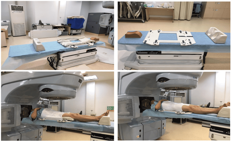
Hình 1. Hai bộ dụng cụ cố định BN xạ trị vùng tiểu khung: bộ dụng cụ cố định có Vac-lock (trái), bộ dụng cụ cố định không có Vac-lock (phải)
For the pelvic immobilization kit using Vac-lock, including: Head-rest pillow, sub-frame fixation table, sub-frame fixation mask, Vac-lock and immobilizer Feet-Fix foot (CIVCO, Orange city, IA). Patients are prepared with an average bladder volume of 350ml before radiation therapy to ensure bladder distension and patient comfort while lying on the treatment table as well as when installing a fixed abdominal mask for the patient. The patient's position is supine on the table and hands are placed in front of the chest, mark the origin fixed on the Vac-lock and write the parameters on the table to fix the subframe, tattoo 3 lead marks as the origin on the patient's skin.
For the pelvic immobilization kit without Vac-lock including: Head-rest pillow, sub-frame fixation table, sub-frame fixation mask and Feet-foot fixation- Fix (CIVCO, Orange city, IA). Patients are prepared with an average bladder volume of 350ml before radiation therapy to ensure bladder distension and patient comfort while lying on the treatment table as well as when installing a fixed abdominal mask for the patient. The patient's position is supine on the table and hands are placed in front of the chest, mark the origin fixed on the Vac-lock and write the parameters on the table to fix the subframe, tattoo 3 lead marks as the origin on the patient's skin.
2.1 2D-KV verification before treatment Before irradiation, all 26 patients were taken daily 2D-KV, 3D-CBCT on Monday and Thursday for subframe VMAT technique. , 2D-KV images are x-ray images of skeletal anatomy. A total of 633 pairs of 2D-KV images and 3D-CBCT image sequences were evaluated for accurate position reconstruction in all 3 AP directions (anterior posterior); SI (top bottom); LR (left right); setting error data due to incorrect position reconstruction is recorded on the OBI (on-board imaging) system. They were compared and contrasted with DRR (Digital reconstruction Radiography) images [ Figure 2] and CT Scans [ Figure 3] to determine the setting error in all 3 directions.

Hình 2. Đánh giá sự tái lập vị trí trên hình ảnh 2D-KV với hình ảnh DRR trước điều trị và sai số cài đặt của sự thay đổi tái lập vị trí chính xác bệnh nhân

Hình 3. Đánh giá sự tái lập vị trí trên hình ảnh 3D-CBCT với hình ảnh CT scans trước điều trị và sai số cài đặt của sự thay đổi tái lập vị trí chính xác bệnh nhân.
2.2 Retrospective study method Based on the comparison of accurate position reconstruction between 2D-KV images with DRR images according to bone landmarks and 3D-CBCT images with CT scans images according to controlled volumes treatment, setting errors due to incorrect repositioning will be recorded prior to treatment sessions in all 3 AP directions; SI; LR. Then it will be matched and Couch Shift to get to the correct treatment location. A total of 633 pairs of 2D-kV and 3D-CBCT images were analyzed and evaluated in all 3 directions to determine:
Average setting error in each direction of all 26 radiotherapy patients for regional tumors Sub-frame. Mean setting error and standard deviation of 13 patients undergoing radiation therapy for pelvic tumors using Vac-lock kits. Mean setting error and standard deviation of 13 patients receiving radiation therapy for pelvic tumors using a kit without Vac-lock Standard deviation for each patient. Standard deviation of the mean setting errors for each patient. Mean of standard deviations for each patient
3. Results and discussion
The change in correct patient positioning during daily radiation was determined and statistically analyzed based on the data of 633 pairs of 2D-kV and 3D-CBCT images in 3 directions AP, SI, respectively. LR:
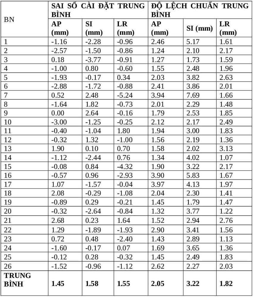
Table 1: Mean setting error of all patients receiving radiation therapy for pelvic tumors.

Table 1 shows the average setting error ± Standard deviation for all 26 patients in the AP, SI, LR directions: (1.45 ± 2.05mm), respectively; (1.58 ± 3.22mm); (1.55 ± 1.82mm).
In 633 initial setting error evaluations, we observed a percentage of initial BN settings with errors exceeding 3mm in AP, SI, LR directions: 7.27%, respectively; 13.28% and 8.14%.

Hình 4: Biểu đồ phân bố sai số cài đặt tái lập vị trí chính xác của bệnh nhân xạ trị vùng tiểu khung.
Reposition error setting using a Vac-lock immobilizer in the subframe:
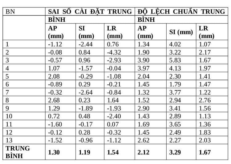
Table 2: Repositioning error of patients undergoing radiotherapy for cervical cancer using the kit Fixed Vac-lock

Average setting error ± standard deviation of 13 patients receiving radiotherapy for pelvic tumors using Vac-lock kits in AP, SI, LR directions are: (1.30 ± 2.12mm), respectively; (1.19 ± 3.29mm); (1.54 ± 1.67mm).

Hình 5: Biểu đồ phân bố sai số cài đặt tái lập vị trí bệnh nhân xạ trị ung thư tiểu khung có sử dụng bộ dụng cụ cố định Vac-lock.
In 385 initial setting error evaluations, we observed a percentage of initial BN settings with errors exceeding 3mm in the AP, SI, LR directions: 3.77%; 6.36%; 3.57%.
Error in 3-way AP; Stable SI and LR and accurate reproducibility of the treatment site are within the allowable treatment limits at the center ≤ 3mm for VMAT radiotherapy. Using a Vac-locked subframe stabilizer allows optimized repositioning of the correct patient position and reduces errors due to patient mobility during irradiation.
Reposition error setting setting error using immobilizer without Vac-lock in subframe :
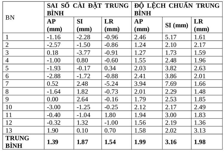
Table 3: Reposition error setting radiotherapy patient for cervical cancer using kit immobilizer without Vac-lock.

Average setting error ± standard deviation of 13 patients receiving radiotherapy for pelvic tumors using the kit without Vac-lock in AP, SI, LR directions is: (1.39 ± 1.99mm, respectively) ; (1.87 ± 3.16mm); (1.54 ± 1.98mm).
In 248 initial setting error evaluations, we observed a proportion of initial BN settings with errors exceeding 3mm in AP, SI, LR directions: 6.85%; 15.52%; 9.68%.
Error in 3-way AP; Stable SI and LR and accurate reproducibility of the treatment site are within the allowable treatment limit at the center ≤ 3mm for VMAT technique radiotherapy. However, the above data table shows that the displacement of treatment repositioning error in the SI (superior-inferior) axis is larger than that of the other two dimensions AP and LR. Therefore, using a Vac-lock-free subframe area fixer may cause more displacement of the repositioning error along the SI (superior-inferior) axis than the other AP and LR axes during projection. radiation to the patient.

Hình 6: Biểu đồ phân bố sai số cài đặt tái lập vị trí bệnh nhân xạ trị ung thư tiểu khung có sử dụng bộ dụng cụ cố định không có Vac-lock.
Comparison of accurate position reproducibility of two Vac-locked and non-Vac-locked kits:
Table 4: Positional accuracy comparison chart of two Vac-locked immobilizer kits -lock and not Vac-lock subframe.
| NHÓM DỤNG CỤ | Z (mm) | Y (mm) | X (mm) |
| VAC-LOCK | 1.30 ± 2.12mm | 1.19 ± 3.29mm | 1.54 ± 1.67mm |
| NON-VAC-LOCK | 1.39 ± 1.99mm | 1.87 ± 3.16mm | 1.54 ± 1.98mm |
The average setting error ± standard deviation when using the fixed kit with Vac-lock in the AP, SI, LR directions is: (1.30 ± 2.12mm), respectively; (1.19 ± 3.29mm); (1.54 ± 1.67mm). The mean ± standard deviation setting error when using the immobilizer kit without Vac-lock in the AP, SI, LR directions is: (1.39 ± 1.99mm), respectively; (1.87 ± 3.16mm); (1.54 ± 1.98mm) respectively. We see that the error when not using Vac-lock is larger than when using Vac-lock in all directions, especially in the SI axis, and the ratio of patient setting errors ≥3mm in the group not using Vac- lock is more frequent than using Vac-lock also in the SI direction.
Table 5: Comparison chart of percentage of correct position reproducibility of two immobilizer kits with Vac-lock and without Vac-lock subframe exceeding the allowable limit ≥3mm.
| NHÓM DỤNG CỤ | Z (tỷ lệ % ≥3mm) | Y (tỷ lệ % ≥3mm) | X (tỷ lệ % ≥3mm) |
| VAC-LOCK | 3.77% | 6.36% | 3.57% |
| NON-VAC-LOCK | 6.85% | 15.52% | 9.68% |
Error in 3-way AP; Stable SI and LR and accurate repositioning of the treatment site are within the allowable treatment limits at the center 3mm for VMAT radiotherapy when both sub-frame immobilizers are selected simultaneously. . However, the data table in Table 5 shows that the displacement error of treatment site re-positioning in the group not using Vac-lock had a non-significantly larger error in repositioning compared to the group using Vac- lock. Therefore, we would prefer to use a Vac-locked pelvic immobilizer while performing treatment of the patient in the pelvic region on a daily basis to optimize accurate repositioning of the patient's treatment site. core.
4. Conclusion
Accurate repositioning of the treatment position when using a Vac-lock immobilizer gives better repositioning results than without using a Vac-lock. Priority is given to the use of a Vac-locked subframe immobilizer to stabilize the patient's position and accurately re-establish the daily treatment position in all AP, SI and LR axes in radiotherapy practice. clinical day at the center.
Please dial HOTLINE for more information or register for an appointment HERE. Download MyVinmec app to make appointments faster and to manage your bookings easily.
REFERENCES
Kneebone A, Gebski V, Hogendroorn N and Turner S. A Randomized Trial Evaluating Rigid Immobilization for Pelvic Irradiation. Int. J. Radiat. Oncol. Biol. Phys., Vol. 56, No. 4, pp. 1105-1111, 2003. Sir MY, Epelman MA and Pollock SM. Stochastic Programming for Off-line Adaptive Radiotherapy. http://www.springerlink.com/content/48m0n0378447w365/ Kasabasic M, Faj D, Simlovic R, Svabi M, Ivkov A and Jurkovic S. Verification of the Patient Positioning in the Bellyboard Pelvic Radiotherapy. Coll. Antropol. 32, Suppl. 2, pp. 211–215, 2008. Kasabasic M, Faj D, Nenad B, Zlatan F and Ilijan T. Implementing of the offline setup correction protocol in pelvic radiotherapy: safety margins and number of images. Radiol.Oncol., Vol.41, No. 1, pp.49-56, 2007. Hunt MA, Schulthesis TE, Desbory GE, Hakki M and Hanks GE. An evaluation of setup uncertainties for patients treated to pelvic sites. Int. J. Radiat. Oncol. Biol. Phys., Vol. 32, No. 1, pp. 227-233, 1995. Lynn V. Immobilizing and Positioning Patients for Radiotherapy. Seminars in Radiat. Oncol., Vol.5, issue 2, pp 100-114, 1995. Hurkmans CW, Remeijer P, Lebesque JV and Mijnheer BJ. Set-up verification using portal imaging; review of current clinical practice. Radiother. Oncol., Vol. 58, pp.105-120, 2001. 27 Clippe S, Sarrut D, Malet C, Miguet S, Ginestet C and Carrie C. Patient setup error Measurement Using 3D intensity-Based Image Registration Techniques. Int. J. Radiat. Oncol. Biol. Phys., Vol. 56, No. 1, pp. 259–265, 2003. Catton C, Lebar L, Warde P, Hao Y, Catton P, Gospondarowicz M, McLean M, and Milosevic M. Improvement in total positioning error for lateral prostatic fields suing a soft immobilization device. Radiother. Oncol., Vol. 44, pp. 265-270, 1997. Creutzeberg CL, Althof VM, de Hoog M, Visser AG, Huizenga H, Wijinmaalen A and Levendag P. A quality control study of the accuracy of patient positioning in irradiation of pelvic fields. Int. J. Radiat. Oncol. Biol. Phys., Vol. 34, No. 3, pp. 697-708, 1996. Suzuki M, Nishimura Y, Nakamatsuk, Okumura M, Hashiba H, Koike R, Kanamori S, Shabibata T. Analysis of interfractional set-up errors and intrafractional organ motions during IMRT for head and neck tumors to define an appropriate planning target volume (PTV)- and planning organs at risk volume (PRV)-margins. Radiother. Oncol., Vol. 78, pp. 283–29, 2006. Thun MJ, Delancey JO, Center MM, Jemal A and Ward EM. The global burden of cancer: priorities for prevention. Carcinogenesis vol.31, No.1, pp.100–110, 2010.


