X-ray technique for straight forearm

This is an automatically translated article.

The article is professionally consulted by MSc, BS. Dang Manh Cuong - Radiologist - Radiology Department - Vinmec Central Park International General Hospital. The doctor has over 18 years of experience in the field of ultrasound - diagnostic imaging.
X-ray technique of straight forearm is applied to diagnose injuries and pathologies in the forearm such as fractures, dislocations, osteomyelitis, all kinds of bone tumors effectively.

1. What is forearm?

The forearm region is the position measured from the three-finger fold below the elbow crease to the furthest crease at the wrist. The forearm region is divided into two sub-regions: the forearm region and the posterior forearm.

2. What is an upright forearm X-ray?


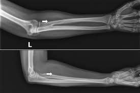
Chụp X quang cẳng tay thẳng nghiêng là kỹ thuật hình ảnh phổ biến dùng để đánh giá các tổn thương ở vùng cẳng tay một cách hiệu quả
Inclination forearm radiograph is a common imaging technique used to effectively evaluate forearm lesions. This technique captures the entire forearm in the supine position, taking both the elbow and wrist joints. Shoot in two planes: Straight and inclined.
Common injuries requiring X-ray of the forearm straight-angle such as: Forearm fracture, forearm dislocation, or inflammatory bone diseases, infections and benign and malignant bone tumors... In which, Two forearm fractures are injuries requiring the application of the most oblique forearm radiographic technique available today.

3. Technical procedure for X-ray of forearm straight-angled

3.1 Preparation Before X-ray of the forearm, the patient will be asked to remove jewelry and metal objects on the body to avoid affecting the imaging technique. If the patient is suspected of being pregnant, immediately notify the doctor to find a safe solution to avoid the negative influence of X-rays on the fetus in the abdomen. The patient may be asked to wear a hospital gown. The patient needs an X-ray order sheet.

Trước khi chụp X quang, bệnh nhân sẽ được yêu cầu cởi đồ trang sức để tránh làm ảnh hưởng đến kỹ thuật chụp
3.2 Steps to perform the oblique forearm X-ray technique The procedure for performing the oblique forearm radiograph technique will be performed by specialist doctors and supported by an radiologist. The patient enters the X-ray room. The doctor receives the X-ray order and compares the part to be scanned with the patient's clinical injury. The doctor explains step by step the procedure to perform the technique for the patient to understand. Electro-optical technician adjusts the mount, the ball is about 1m away. The doctor instructs the patient to sit next to the X-ray table, the patient's elbow to be taken is extended, the palm is facing up, the back of the forearm is placed close to the film. Tilt position with elbow flexing the outer edge of the forearm close to the film. The technique of adjusting the center ray to the middle of the forearm. Paste the letter (F) (T) corresponding to the patient's hand on the digitizer plate. Save the patient's name and age on the computer and then select the program corresponding to the part to be taken. Factor adjustment technique (48kv; 1.2mAs) The doctor asks the patient to hold their breath for a short time to get the clearest image. Close the door of the imaging room and give the X-ray. After taking the picture, the patient was directed to the room to wait for the results. The technician adjusts the contrast and checks the balance of the images on the film. Print film. 3.3 Evaluation of the scan results Passed results: Taken above and below one joint, at least one joint closest to the lesion. In the upright-tilt position, the entire forearm bones are obtained. X-ray quality: Sharp, high contrast. X-ray film with patient's name, age, mark (F) (T), date and year taken. The doctor evaluates the lesion based on the obtained computer images. The doctor can give professional advice to the patient if required. 3.4 Complications and management The technique of taking an upright forearm radiograph without complications.
However, in some cases, the technique will have to be repeated because the patient does not keep still during the film, so the results are not clear, the lesions are not clearly visible.

Please dial HOTLINE for more information or register for an appointment HERE. Download MyVinmec app to make appointments faster and to manage your bookings easily.

SEE MORE
Treatments for forearm and arm fractures Signs of forearm and arm fractures Care and recovery after fractures
Share
Patients Stories