This is an automatically translated article.
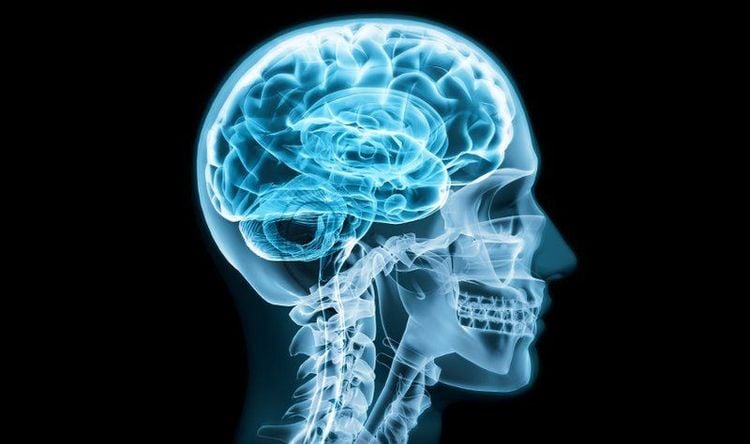
This article is professionally consulted by Master, Resident Doctor Tran Duc Tuan - Department of Diagnostic Imaging - Vinmec Central Park International General Hospital.Craniofacial X-rays are X-ray images of the bones that surround the brain, face, nose, and sinuses. Craniofacial X-rays are often aimed at quickly finding signs of trauma, thereby helping to guide the initial treatment steps as well as the means of subsequent imaging studies.
1. Anatomy of the craniofacial bones
The skull has a structure like a sealed box, which is an important part to help protect the brain inside. Beneath the skull forward are the facial bones, which help shape the structure of the face and the entire jaw and mouth area.
All the bones that make up the skull are attached to each other by immovable joints, with the exception of the jawbone, which is attached via a movable joint.
The structure of the skull is made up of 8 bones, including the frontal bone, the parietal bone (consisting of the two bones on the sides), the temporal bone (consisting of the two bones on the sides), the ethmoid bone, the sphenoid bone, and the occipital bone.
Meanwhile, the skeleton of the face is composed of more bones, smaller in size and more detailed, which helps to give a sophisticated shape to the face, including the shape of the eyes, nose and mouth.
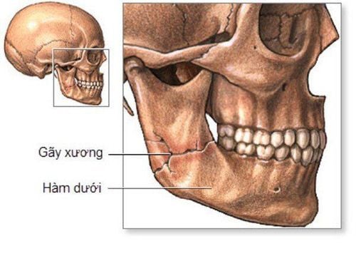
Xương hàm là xương duy nhất trên hộp sọ được gắn với nhau qua một khớp động
2. What is a craniofacial X-ray?
X-rays, which use beams of invisible electromagnetic energy to create images of any part of the body, have long been commonly used in medicine to diagnose and treat diseases. For the whole skull, a standard craniofacial x-ray has also been performed for many reasons such as tumor diagnosis, infection, foreign body, or bone trauma.
The principle of X-rays is to use X-rays to pass through body tissues into specially treated plates (similar to camera film). As the body undergoes X-rays, different parts of the body allow different beams of X-rays to pass through. Images are produced in light and dark levels, depending on how much x-rays penetrate the tissues, the stronger the structure, the clearer it appears on the film, and vice versa.
Soft tissues in the body (such as blood, skin, fat, and muscle) allow most x-rays to pass and appear dark gray on the film. A bone or a tumor, which is denser than soft tissue, allows some X-rays to pass through and appears white on the X-ray. In a fracture, the X-ray beam passes through the broken area and appears as a single bone. Dark lines in white bones.
Today, with the development of technology, computers and digital media are now more commonly used in place of film, helping to produce sharp and detailed high-resolution images. At the same time, although facial X-rays are not used as often as they used to be, due to the trend toward newer technologies such as CT scans and cranial MRI, cranial radiographs are still the most useful. Determined to find signs of craniofacial fracture as quickly as possible and detect other conditions of the skull and brain, sinuses.

Phương pháp chẩn đoán hình ảnh hiện đại MRI sọ não
3. When to take a cranial X-ray?
Cranial X-ray is indicated to diagnose skull fractures, birth defects, infections, foreign bodies, pituitary tumors as well as some metabolic and endocrine disorders that cause skull defects . In addition, skull X-rays can also be used to look for tumors, examine the sinuses, and detect calcifications in the brain.
Although cranial X-rays are associated with a risk of radiation exposure, this is only to a small extent and the benefits outweigh the potential risks. However, special populations such as pregnant women and children, who are more sensitive to the risks associated with X-rays, should be carefully considered when ordering cranial x-rays.
4. How is the skull X-ray taken?
When a cranial X-ray is ordered, a radiologist or radiologist will explain to you how it is done. In general, however, cranial X-rays do not require special preparation, including fasting.

Người bệnh không cần nhịn ăn trước khi tiến hành chụp X-quang sọ não
X-rays can be taken during your stay in the hospital or as an outpatient, which means you can go home immediately after the scan. No matter where a cranial X-ray is taken and with what specific indications, a basic procedure for a skull X-ray should be as follows:
You will be asked to change your clothes, remove all jewelry sets, hairpins, eyeglasses, hearing aids, or other metal objects that can interfere with the X-ray. Wear a special gown.
You are properly positioned for the cranial X-ray and need to focus on remaining in a certain position for a few minutes for the technician to release the X-ray.
If a cranial X-ray is needed to When looking for an intracranial injury, special care should be taken to prevent further injury, eg using a neck brace if a cervical spine fracture is suspected.
The technician may ask to be repositioned for X-ray examination from different angles. It is important that you remain completely still while shooting.
Although the X-ray procedure is painless, moving the body parts to be examined can cause some discomfort, especially in the case of trauma or a recent invasive procedure like surgery. The patient should be equipped with the best means of reinforcement and the radiologist should also complete the X-ray as quickly as possible to minimize any discomfort or pain for the patient.
Chụp X-quang sọ không gây đau đớn hay khó chịu cho người bệnh
When the X-ray is finished, you can go home and do your normal activities, except for injuries that may limit your movement. The results of the film will be analyzed by a radiologist and sent back to the designated doctor for diagnosis and treatment. In some cases of suspected pathology, you may need further investigation with more specialized imaging facilities such as CT or MRI of the brain.
In summary, X-ray has always been the leading basic imaging tool to investigate bone pathologies. For craniofacial, X-ray helps to detect early signs of fracture in trauma as well as some maxillofacial pathologies. It is still considered a useful tool in emergency situations and especially where CT and MRI of the brain are not available.
Vinmec International General Hospital is a quality medical facility of Vietnam, equipped with modern medical equipment, imported from the US, Japan, the Netherlands, Korea... to help diagnose and treatment is most effective. The doctors are well-trained, specialized from abroad, rich in experience. Treatment methods are updated regularly, on par with major hospitals.
Master, Doctor Tran Duc Tuan is well-trained in the country and many centers have prestigious medical background in the world such as: Australia, Singapore, Thailand... The doctor has many years of experience in the field of medicine. imaging and endovascular and extravascular interventions. Currently, the doctor is working at the Department of Diagnostic Imaging and Interventional Vascular Unit - Vinmec Central Park International General Hospital.
To register for examination and treatment at Vinmec International General Hospital, you can contact the nationwide Vinmec Health System Hotline, or register online HERE.
References: radiopaedia.org , hopkinsmedicine.org , mountsinai.org


