
This is an automatically translated article.
The article was professionally consulted by resident Doctor Nguyen Quynh Giang - Department of Diagnostic Imaging and Nuclear Medicine - Vinmec Times City International Hospital. Dr. Giang has many years of experience in the field of diagnostic imaging, especially in the field of multi-segment computed tomography, magnetic resonance.X-ray is the most important laboratory test in the diagnosis of lung or bone disease. Chest X-ray is a routine test in the examination and treatment of many diseases, especially those related to the respiratory tract.
Today, despite modern technology, there are many advanced and specialized imaging tools with high accuracy, but X-ray has never been underestimated in the diagnostic criteria. The article will provide information about basic chest radiographs as well as basic chest X-ray reading steps.
1. What is a chest X-ray? Posture during chest x-ray?
Chest radiograph is a diagnostic imaging technique used to evaluate the condition of the patient's chest, its components, and adjacent structures. Chest X-ray is the most commonly taken film in medicine, this method is most used by doctors to evaluate, help detect abnormal symptoms of chest X-ray films, thereby helping clinicians to make a preliminary diagnosis.Like other X-ray methods, a chest X-ray uses radiation to form X-rays from which an image of the chest is taken. The average radiation dose for adults is about 0.02 mSv (2 mrem) for a straight chest film (PA or posterior-anterior) and 0.08 mSv (8 mrem) for a lateral film (LL or latero-lateral).
Detailed instructions on how to detect abnormalities on chest x-ray can help clinicians make a preliminary diagnosis.
These are really valuable sharing with not only radiologists but also help clinicians improve their knowledge and skills in disease diagnosis.
There are 4 main positions when taking X-rays:
Back - front Tilted Front - back Lying - side
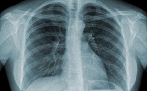
X quang ngực là phương pháp kỹ thuật dùng để chẩn đoán hình ảnh nhằm đánh giá tình trạng của ngực
2. When should a chest X-ray be performed?
Patients with chest X-ray are indicated when the doctor suspects or needs to evaluate the patient's condition related to the lungs, bones:Pneumothorax Pneumothorax Interstitial lung disease Heart failure Fracture Fracture Diaphragmatic hernia Benefits of X-ray film:
Simple shooting technique, easy to implement. Low cost. Time to have short film shooting results. The disadvantage of this method is that the accuracy of the radiograph results or not completely depends on the skill of the photographer, the cooperation of the patient and the doctor's experience and ability to read the results.
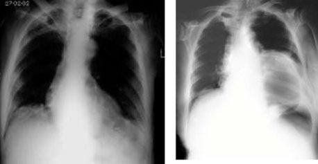
Bệnh nhân có chỉ định chụp X quang ngực khi bác sỹ nghi ngờ hoặc cần đánh giá tình trạng của bệnh nhân liên quan tới phổi
3. Steps to read chest X-ray
Recheck patient information, X-ray date. Attention should be paid to the shooting position: straight, at the bed, tilted, slanted, lying on the side. Viewing film markings Viewing film quality: Is it too light or too dark, inhale deeply enough? Real estate good? Is the posture right? Instructions for reading chest radiographsChest radiograph results are normal when:
Thoracic bones:
The sternum and thoracic spine are usually not visible on straight films, only on lateral radiographs.
Ribs : Determined on straight film.
Posterior: Going from top to bottom, inside to outside. Anterior bow: Going from top to bottom, from outside to inside. The cartilage is not contrasted, so the lesion cannot be identified. Ribs 11, 12 can only be seen on urography. Muscles and soft parts of the chest wall:
Pectoralis major, sternocleidomastoid muscle. Silhouettes of women's breasts and nipples. These parts overlap the background and periphery of the two lung fields, reducing the brightness of the lungs.
Lung parenchyma and hilum:
Two lung fields that are black on radiographs are called bright images. The umbilicus has a white, so-called opacity, originating from either side of the heart margin, in the shape of a tree root. The hilum is formed by the pulmonary artery and the bronchi. The left lung hilum is usually 1-2 cm higher than the right lung hilum. The hilum of the lung divides gradually to the periphery into the pulmonary ridge. When about 1 cm from the chest wall, the pulmonary ridges are no longer visible. Cardiac and mediastinal shadow:
Normal in left skewed position and <1⁄2 transverse thoracic diameter. Trachea and main bronchus
Visible on high-voltage radiographs. The right diaphragm is about 1-2 cm higher than the left diaphragm. Just below the left diaphragmatic arch is the gastric air sac.

Chụp X quang là kỹ thuật phổ biến mà bất cứ bệnh viện nào cũng có
Separation of the waste field On straight film:
Horizontally:
Apical region: From the upper border of the 2nd anterior arch and upwards. Umbilical region: From the superior border of the 2nd rib anterior to the 4th anterior arch. Vertical:
Center zone: Along the mid-clavicle point inward. Peripheral region: Along the midpoint of the collarbone outward. According to radiographic anatomical landmarks:
The region above the clavicle. Subclavian region. Horizontal flank angle on both sides. The central angle of the diaphragm on both sides. Lung hilum area. On slant film:
Lungs are divided into lobes and lobes. The right lung has 3 lobes, upper, middle and lower. The left lung has 2 lobes, upper and lower. Each lung is divided into 10 lobes. Mediastinal zoning
Straight plane :
The mediastinum is a watermark located between the two lung fields, bounded by the bilateral mediastinal pleura. Divided into 3 floors (upper, middle and lower) by two planes through the superior border of the aortic arch and through the inferior border of the tracheobronchial junction. Inclined plane:
Divided according to Felson's diagram:
Anterior mediastinum: From the posterior surface of the sternum to the anterior border of the trachea (or the posterior border of the heart). Mediastinum: The anterior mediastinum follows the anterior border of the thoracic spine about 1cm. Posterior mediastinum: Next to the middle mediastinum to the end of the rib trough. X-ray is a common technique that any hospital has. Vinmec International General Hospital uses X-ray techniques to diagnose diseases. With a team of highly qualified doctors, modern medical equipment and machinery, meeting international standards, when in need of examination or treatment, please contact the hotline number to receive the earliest advice and support. maybe.
Please dial HOTLINE for more information or register for an appointment HERE. Download MyVinmec app to make appointments faster and to manage your bookings easily.