This is an automatically translated article.
The article was professionally consulted by Dr. Nguyen Anh Tu - Doctor of Obstetric Ultrasound - Prenatal Diagnosis - Obstetrics Department - Vinmec Hai Phong International General Hospital.Thanks to contrast material, the esophagus, stomach, and duodenum will be clearly highlighted on the X-ray film, through the results of X-ray of the esophagus - stomach, doctors will more accurately diagnose the condition. condition and more effective treatment regimens. X-ray technique of the esophagus - stomach is now very indicated in the diagnosis and treatment.
1. X-ray of esophagus - stomach when?
X-ray technique will use invisible electrical energy beams to create images of tissues in internal organs, bones of the body on x-ray. X-ray is one of the imaging techniques used quite commonly in the diagnosis and treatment of diseases, in fact, this is a procedure that uses X-rays to reconstruct images of organs in the body. The doctor makes a diagnosis. During an X-ray, X-rays pass through the patient's body and are recorded on a shield behind the patient from which an image is produced.
Esophageal-stomach X-ray technique is performed on a television brightening X-ray machine with a compression set used for gastric and intestinal imaging. Currently, thanks to the advantages of flexible endoscopic technique combined with biopsy in the diagnosis of gastroduodenal diseases, the reliability is quite high, along with the development of imaging techniques such as microscopic tomography. Computer, ultrasound, magnetic resonance ... so the role of X-ray of the esophagus - stomach is limited in some cases, such as assessing the extent of lesions in the stomach and duodenum, in those cases not Endoscopy is possible, or the patient does not cooperate with endoscopy.

Người bệnh có tổn thương ở dạ dày nên chụp X-quang
2. How does the oesophageal-stomach X-ray procedure take place?
The procedure for taking X-ray of the esophagus - stomach will take place as follows:
Step 1: The technician prepares the means and supplies for the scan.
Step 2: The patient takes gastric contrast agent
Step 3: Take a series of films (two or three) in the same position, simultaneously taking many different positions, which is important in the assessment of function of each region.
Mucosal scan: Patient lying supine and slightly left front: swallow 60ml Barite. The table is slightly sloping, barique is spread on the back. Turn the patient into the right-back position, turn back and forth so that the drug adheres to the frontal mucosa. Take 2 movies: one front, one back. Take full medicine: Standing table, give the patient 150-200ml to drink: take 2 films while the patient swallows, take the junction of the esophagus, the cardia, the aneurysm in the right anterior position. When the stomach is full, take one straight, anterior right and one 24x30cm slant film. Turn the table horizontally, the patient lies on his back, take a 24x30cm film. The patient lies on his stomach and right anteriorly to separate the duodenal frame from the duodenal bulb. Take a series of 4 images on 30x40cm film. With the digital system can be smaller size 18x24 cm, or 35x43 cm divided into 4 images. Take imaging to look for reflux esophagitis, compress when necessary. Double contrast imaging: Consists of 2 main phases: lying on the back, taking the back and lying on the stomach, capturing the front. Esophageal-gastric x-ray technique is a non-invasive technique, the risk of complications is very low. However, to ensure safety, it is absolutely forbidden to take Baryt contrast in patients with suspected perforation of a hollow viscera or intestinal obstruction.
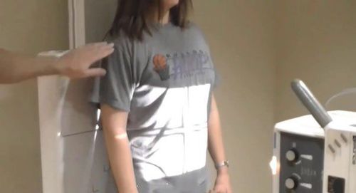
Kỹ thuật viên hướng dẫn người bệnh chuẩn bị tư thế chụp
3. Note when taking X-ray of esophagus - stomach
Patients should take X-ray of the esophagus - stomach in the morning, need to fast (not eat, not drink) and do not smoke within 8 hours before the scan. In particular, the patient should not take drugs containing contrast medium within 3 days before the scan.
X-ray techniques as well as many other medical examination and treatment techniques at Vinmec International General Hospital are conducted by a team of highly qualified and experienced medical doctors; the system of advanced and modern machinery should significantly reduce radiation dose with the best image and always be updated according to medical progress in the world; Especially, professional service quality will help customers have the most comfortable and secure experience when visiting Vinmec. In addition, at Vinmec International General Hospital, there is a package of screening and early detection of cancers of the gastrointestinal tract (esophagus - stomach - colon) combined with clinical and subclinical examination to bring about key results. as accurate as possible.
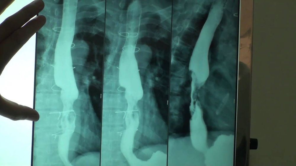
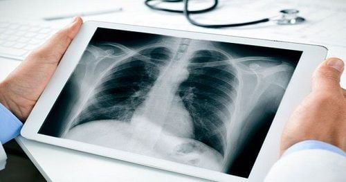
Hình ảnh X-quang thực quản
When screening for gastrointestinal cancer at Vinmec, you will receive:
Gastrointestinal specialty examination with an oncologist (by appointment). Gastroscopy and colonoscopy with an NBI endoscope under anesthesia. Total peripheral blood cytology (by laser counter). Automated prothrombin time test. Automated thrombin time test. Activated Partial Thromboplastin Time (APTT) test using an automated machine. General abdominal ultrasound To register for screening and treatment of gastrointestinal diseases at Vinmec International General Hospital, you can contact Vinmec Health System nationwide, or register for an online examination. HERE .
SEE MORE
Relationship between X-rays and Pregnancy In what case is X-ray of the uterus and fallopian tubes (HSG scan) done? Mammomat Inspiration X-ray machine - an effective "assistant" in the diagnosis and treatment of breast tumors


