This is an automatically translated article.
The article is professionally consulted by Master, Doctor Nguyen Hong Hai - Radiologist - Department of Diagnostic Imaging and Nuclear Medicine - Vinmec Times City International Hospital
The bladder is the place where urine is transported from the kidneys to the kidneys and then out of the body through the urethra. Any problem with the bladder affects its function and ability to function. Bladder ultrasound is an effective imaging technique to help identify and diagnose diseases that occur in this area early.
1. Bladder characteristics
The bladder is a hollow organ whose shape, size and position varies according to the amount of urine it contains. The bladder receives urine from the 2 kidneys and then excretes the urine through the urethra.
The average bladder can hold up to 500ml of urine without being too full. When the bladder normally holds 250-350ml of urine, we feel the urge to urinate.
The bladder is located below the peritoneum, when emptying the bladder will lie completely in the anterior part of the pelvis. In front of the bladder is the pubic bone, behind is the rectum and genital organs, below the bladder is the pelvic diaphragm.

Bàng quang là một tạng rỗng mà hình dạng, kích thước và vị trí thay đổi tùy theo lượng nước tiểu chứa bên trong nó.
2. The role of bladder ultrasound
When experiencing problems related to urinary disorders, bladder ultrasound is the first method used to examine and diagnose the condition. Bladder ultrasound is a simple and highly effective measure, with mild patients often only needing this method.
Bladder ultrasound plays a role:
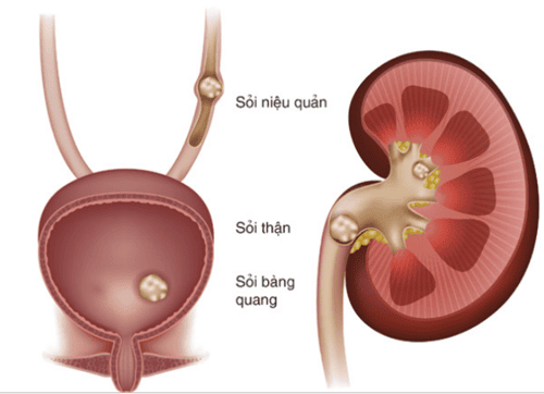
Detect tumors, assess the extent of invasion. Recognizing congenital malformations of the bladder, bladder diverticulum.... Evaluation of urinary retention; Evaluation of bladder stones, lower ureteral stones. Evaluation of bladder injury, blood in the bladder. Detection of diseases: prostatitis, urethral stones, cystitis, overactive bladder, bladder cancer,... From the information above, the doctor will soon determine the condition that the patient is experiencing. right.
For bladder ultrasound, it can be accessed through 4 routes:
The suprapubic line. The way through the perineum. Vaginal tract. Rectal tract. Technically, the doctor will use a specialized transducer that emits ultrasound waves to examine the entire abdominal cavity of the patient. This technique is painless and does not cause damage to the bladder. Before performing a bladder ultrasound, the doctor will ask the patient to drink a lot of water and hold back urine to keep the bladder full.

Trước khi tiến hành siêu âm bàng quang, bác sĩ sẽ yêu cầu người bệnh uống nhiều nước và nhịn tiểu để bàng quang căng lên.
3. Bladder ultrasound image
The bladder has a triangular tetrahedron with 4 faces: the upper, the posterior and 2 lower sides.
Upper: Covered by the peritoneum, convex when the bladder is full and concave when the bladder is empty, giving the bladder a Y or T shape on a vertical plan. 2 inferior lateral faces: Meet anteriorly by a rounded margin, sometimes called anterior, resting against the pelvic diaphragm. Inferior posterior: Flat, sometimes convex, also known as the base (floor) of the bladder. And the upper part of the bladder bottom is covered by the peritoneum. The shape of the bladder will change depending on the state of urine storage. A normal bladder ultrasound image will have the following features:
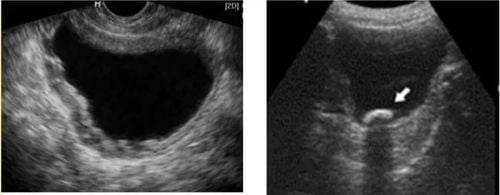
When the bladder is full, the cross section will be square or rectangular with rounded corners, and the longitudinal section will have a triangle shape with rounded corners. Normal bladder wall smooth border, homogenous hyperechoic, 1-3mm thick when bladder distension, 5mm thick when bladder less urine. The triangle of the bladder is made up of 2 holes of the ureter and the opening of the urethra, 4-5mm thick, less negative than other regions. Bladder hole: Easier to see in longitudinal section, inverted caret shape. Ureteral orifice: At the point of emptying into the bladder ureter, up to 2-3mm thick. Conversely, if the bladder is abnormal, the ultrasound image will show the following conditions:
Change in bladder wall. Unbalanced bladder. In or out of the bladder wall appears a cystic structure. Solid structures in or at the base of the bladder (stones, blood clots, tumors, ureteral cysts, ...)

Hình thể bàng quang sẽ thay đổi tùy theo tình trạng chứa nước tiểu.
4. Why is it important to hold urine before bladder ultrasound?
To check for problems related to the urinary system, bladder ultrasound is often used, whereby a full bladder will reflect a clearer and fuller ultrasound image than an empty bladder (no bladder). contain urine).
Thus, before performing a bladder ultrasound, the patient should drink a lot of water and hold urine to increase the visibility of the image, helping to give a more accurate diagnosis. For other parts of the body that need an ultrasound, your doctor will guide you on whether or not you need to have the procedure.
Bladder ultrasound is a way to help detect and diagnose some diseases in the urinary system quickly and effectively. If you suspect you have the disease, you can come to Vinmec International General Hospital System. As one of the leading prestigious hospitals in the country, Vinmec uses the most modern generations of color ultrasound machines today in examining and treating patients. One of them is GE Healthcarecar's Logig E9 ultrasound machine with full options, HD resolution probes for clear images, accurate assessment of lesions. In addition, a team of experienced doctors and nurses will greatly assist in the diagnosis and early detection of abnormal signs of the body in order to provide timely treatment for urinary system pathologies.
Please dial HOTLINE for more information or register for an appointment HERE. Download MyVinmec app to make appointments faster and to manage your bookings easily.