This is an automatically translated article.
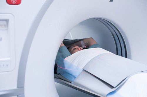
The article is professionally consulted by Master, Doctor Nguyen Thanh Nam - Radiologist - Department of Diagnostic Imaging - Vinmec Danang International General Hospital. The doctor has over 10 years of experience in the fields of Diagnostic Imaging.To diagnose meningiomas most accurately, doctors often use the method of brain MRI. This is an imaging method that is highly appreciated by professional doctors and is often indicated when the patient has clinical symptoms suggestive of the above because it can provide the clearest and most detailed images. on brain parenchymal composition.
1. What is a meningioma?
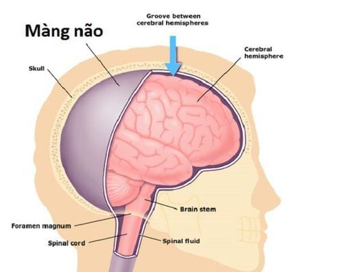
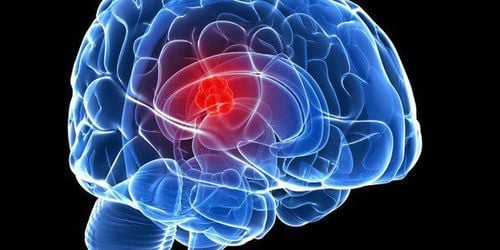
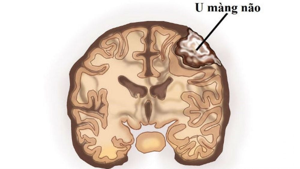
Meningiomas are the most common intracranial tumors, and most of these tumors are benign. Meningiomas are derived from capsular cells in the arachnoid membrane (this is one of the three layers of meninges that surround the brain and spinal cord. Most are benign, but in some cases the control can grow to a very large size and in some special locations can cause neurological dysfunction, thereby affecting the patient's life.Most people develop only one meningioma. veins of the brain and the floor of the skull - these are the places where there are many capsular cells in the arachnoid membrane Some types of meningioma are classified by location as follows:
Cavernous meningiomas; Meningiomas ; Meningiomas of the brain hemispheres; Meningiomas in the orbits Meninges in the ventricles; Meninges in the olfactory grooves; Meningioma of the posterior fossa; Meningioma of the meninges of the cerebellum; Meninges of the sphenoid; Meningioma spine; Meningiomas o superior cistern., saddle: near the base of the skull, the pituitary gland...
2. Meningioma symptoms
Meningiomas usually grow very slowly and cause few noticeable symptoms until they are quite large. Symptoms of meningioma depend on the size and location of the tumor as follows:Tumor location in the crescent of the brain: In this position, the tumor can cause the patient to have impaired neurological function. . Where the tumor is located in the midline, it may cause lower extremity weakness, paresthesia or convulsions; Tumor location in the hemisphere: The tumor may cause seizures, headaches, or neurological deficits for patients; Tumor location in the sphenoid bone: In the sphenoid bone, the tumor may affect vision, loss of sensation in the face, facial paresthesia or convulsions; Olfactory groove tumor: Because it is in the olfactory groove, the tumor compresses the olfactory nerve, thereby causing the patient to lose the sense of smell. In case the tumor grows larger, it may affect vision because the optic nerve is compressed by the tumor; Tumor location in the suprapubic basin: Affects vision due to tumor compression of the optic nerve/optical nerve; Posterior fossa: Facial symptoms or hearing loss due to compression of cranial nerves, unsteady gait and loss of coordination; Tumor location in the ventricles: If the tumor is in this location, it will lead to blockage of cerebrospinal fluid drainage, headache, dizziness or cause neuropsychiatric dysfunction; Tumor location in the orbit: If the tumor is in the orbit, it will cause increased orbital pressure, protrusion or worse, the risk of vision loss; Spine: Back pain, limb pain caused by nerve compression tumor.

3. The role of MRI method in diagnosing meningioma
Diagnostic methods for meningiomas are often difficult, because most meningiomas are slow growing and mostly only seen in adults, so the symptoms of the disease can often be unclear, making patients and the doctor ignored it.Besides, there are a few symptoms associated with meningiomas that can also be caused by other conditions. Therefore, the usual diagnosis will not be accurate.
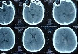
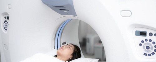
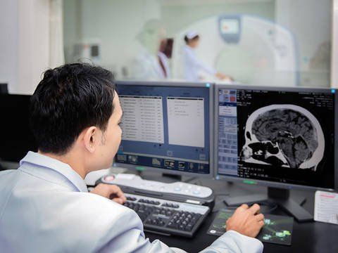
Therefore, in order to diagnose meningiomas most accurately, doctors often use the brain MRI method. This is an imaging method that is highly appreciated by professional doctors and is often indicated when the patient has clinical symptoms suggestive of the above. This method is highly appreciated in the diagnosis of meningiomas because:
Cranial MRI can provide the clearest and most detailed images of brain parenchymal components that cannot be seen on X-rays. clear. Besides, the MRI method to diagnose meningiomas is also useful in finding the cause of abnormal symptoms such as: muscle weakness, muscle paralysis, dizziness, prolonged headache, seizures.... This method can also help detect brain diseases such as hemorrhage, tumor, edema; Brain damage occurs due to trauma or stroke, especially early detection of very small cerebral infarctions without clinical abnormalities. Therefore, doctors recommend that in cases of persistent headache, it can be further evaluated by exploration by means of MRI to diagnose meningiomas or CT tomography.
4. Meningiomas treatment
The treatment for a meningioma will depend on the location and size of the tumour. The treatment methods usually chosen by the doctor are surgery and monitoring the progress of the disease.The surgical methods often depend on the tumor location such as:
Classic craniotomy; Laparoscopic surgery through the sphenoid sinus; Transventricular endoscopic surgery; Minimally invasive surgery to open the keyhole skull cap. The monitoring method is indicated in some cases such as:
Meningeal tumor is small and has no clinical symptoms, only discovered by chance; Tumor is located in an important functional area of the human body such as: tumor in the motor area, around a large blood vessel such as the internal carotid artery or at the location of the large venous sinus; The tumor was followed for a follow-up period and did not tend to increase in size significantly; Meningiomas that have been operated on but have a residual remnant or recur in locations where surgery is difficult to perform. To ensure safety and have an effective treatment regimen, patients need to go to a reputable hospital to conduct examination and treatment as soon as there are signs of meningioma. Currently, Vinmec International General Hospital is one of the leading prestigious hospitals in the country, trusted by a large number of patients for medical examination and treatment. Not only the physical system, modern equipment: 6 ultrasound rooms, 4 DR X-ray rooms (1 full-axis machine, 1 light machine, 1 general machine and 1 mammography machine) , 2 DR portable X-ray machines, 2 multi-row CT scanner rooms (1 128 rows and 1 16 arrays), 2 Magnetic resonance imaging rooms (1 3 Tesla and 1 1.5 Tesla), 1 room for 2 levels of interventional angiography and 1 room to measure bone mineral density.... Vinmec is also the place to gather a team of experienced doctors and nurses who will greatly assist in diagnosis and detection. early signs of abnormality in the patient's body. In particular, with a space designed according to 5-star hotel standards, Vinmec ensures to bring the patient the most comfort, friendliness and peace of mind.
Please dial HOTLINE for more information or register for an appointment HERE. Download MyVinmec app to make appointments faster and to manage your bookings easily.


