This is an automatically translated article.
The meninges are composed of a layer of connective tissue that covers the brain and spinal cord. The meninges consist of 3 different membranes, each of which plays an important role in the functioning of the central nervous system.1. Function of the meninges
Meninges have an important function in protecting and supporting the central nervous system (CNS). The meninges connect the brain and spinal cord to the skull and spinal canal. Accordingly, the meninges act as a protective barrier, helping the sensitive organs of the CNS to avoid injury. It also contains a large number of blood vessels, which supply blood to the cells of the CNS.
An important function of the meninges is to produce cerebrospinal fluid, which fills the cavities of the ventricles that surround the brain and spinal cord. Cerebrospinal fluid protects and nourishes CNS tissue by acting similarly to a shock absorber, in addition to helping to circulate nutrients and remove waste products.
2. Anatomical meninges include what?
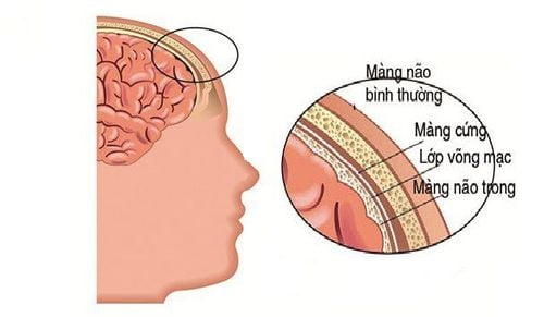
Cấu tạo giải phẫu của màng não
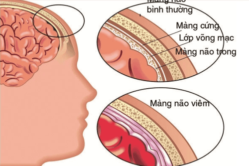
The human brain is enclosed in an outer shell called the scapula. The cranial bones have the function of protecting the brain from damage by combining with the protective bones of the face, forming the skull structure. The separation between the skull and the brain is the meninges. So what is the meninges? Meningeal anatomy will help us answer this question. The meninges consist of three layers of tissue, which cover and protect the brain and spinal cord. From the outside to the inside, the meninges are: dura, arachnoid, and soft membrane, respectively.
2.1. The dura is the outermost layer, connecting the meninges to the skull and spine. The dura is composed of tough, fibrous connective tissue. The dural structure surrounding the brain consists of two layers:
Basement membrane (located outside): responsible for connecting the dura mater to the skull and covering the meninges, with a fairly solid structure. Meningeal layer (inside): considered the actual dura. Between these two layers are specialized channels, known as the dural venous sinuses. These veins carry blood from the brain to the internal veins, which then return to the heart. The meninges also form dural folds, which divide the cranial cavity into different compartments, perform supporting functions, and contain different sections of the brain.
The dura mater forms a tubular sheath that covers the cranial nerves in the skull. On the other hand, the dura mater of the spine is composed only of the meninges, not containing the basement membrane.
2.2. The arachnoid membrane The arachnoid membrane is the second layer of the anatomical structure of the meninges, connecting the dura mater and the soft membrane. The arachnoid membrane covers the brain and spinal cord loosely and has a characteristic arachnoid-like appearance. The subarachnoid space is a route for blood vessels and nerves to travel through the brain and collects cerebrospinal fluid from the fourth ventricle.
2.3. Soft membrane The soft membrane is a thin inner membrane that is in direct contact and closely covers the cerebral cortex and spinal cord. The vascular supply to the soft membranes is very rich, which helps to transport nutrients to the nervous system.
It also contains the choroid plexus, which is a network of capillaries and epithelium (special epithelial tissue) that produces cerebrospinal fluid. The soft membrane covering the spinal cord consists of two layers, the outer layer is composed of collagen fibers, and the inner layer surrounds the entire spinal cord.
Because the function of the meninges is so important to the central nervous system, problems with the meninges are mostly serious.
3. Diseases related to the meninges
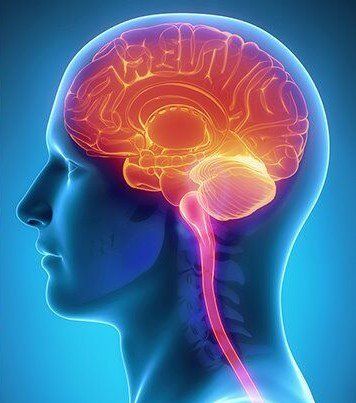
Viêm màng não là một bệnh lý nguy hiểm của màng não
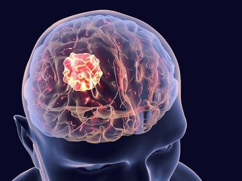
3.1. Meningiomas Meningiomas are tumors that develop in the lining of the brain. The disease begins in the arachnoid layer, putting pressure on the brain and spinal cord as the tumor grows. Most meningiomas are benign and grow quite slowly. However, some meningiomas progress very quickly and become cancerous. Meningiomas can grow into a very large tumor and treatment is usually surgical removal.
3.2. Meningitis Meningitis is a dangerous disease of the meninges. Meningitis is usually caused by an infection of the cerebrospinal fluid. Pathogens are bacteria, viruses, and fungi. Meningitis can lead to brain damage, seizures, and even death if the patient is not treated promptly.
3.3. Hematoma Damage to a blood vessel in the brain can cause blood to accumulate in the brain cavities and brain tissue, forming a hematoma. A hematoma in the brain leads to inflammation, swelling, and possibly damage to brain tissue. The two most common types of hematomas involving the meninges are epidural hematomas and subdural hematomas.
Epidural hematoma occurs between the dura mater and the skull, originates from damage to the arteries or the venous sinuses, caused by severe trauma to the head area.
Meanwhile, a subdural hematoma occurs between the dura and the arachnoid, caused by a head injury, resulting in a ruptured vein. A subdural hematoma is likely to be an emergency emergency, progresses rapidly, or can be slow over a long period of time.
Understanding the structure of the meninges and diseases related to the meninges helps us to be proactive in preventing and dealing with them.
Please dial HOTLINE for more information or register for an appointment HERE. Download MyVinmec app to make appointments faster and to manage your bookings easily.


