This is an automatically translated article.
Heart valve repair surgery or heart valve replacement is a method indicated in cases where the patient's valvular system is damaged causing valve regurgitation, severe stenosis or both stenosis and regurgitation causing many complications for the patient. .1. How does the heart valve system work?
A normal heart has 4 main types of heart valves, including:
Tricuspid valve: The valve located between the right atrium and right ventricle, has the role of bringing blood one way from the right atrium to the right ventricle. Pulmonary valve: Consists of 3 small bird's nest-shaped valves, located between the right ventricle and the pulmonary artery. Blood flow from the right ventricle through the pulmonary valve into the pulmonary artery carries blood to the lungs for the purpose of oxygen exchange. Mitral valve: The valve located between the left ventricle and the left atrium, has the role of transporting blood in one direction from the left atrium to the left ventricle. Aortic valve: Located between the left ventricle and aorta, blood flows from the left ventricle through the aortic valve into the aorta, bringing blood to the whole body. The heart valves have a very important role in coordinating the direction of blood flow in a certain direction, controlling the flow of blood through the heart by opening and closing each time the atria and ventricles contract. The opening and closing functions of such valves are controlled by the pressure difference between the chambers of the heart.
Common heart valve diseases include: Heart valve regurgitation (mitral valve regurgitation, aortic regurgitation); valvular stenosis (most commonly mitral stenosis, aortic stenosis); There may also be some cases of both stenosis and regurgitation in one valve, or multiple valvular stenosis (mitral regurgitation with aortic regurgitation). When the valve is narrowed (the leaflets do not open completely), blood cannot pass through the valve easily, causing circulatory stagnation upstream and ischemia downstream, causing the heart chambers to dilate and heart failure. When the valve is open (the valve leaf does not close tightly), the blood does not flow in one direction, but partially flows back upstream, so the heart has to work harder, which leads to thickening of the heart muscle, dilated heart chambers, and heart failure. .
If compared with mitral and aortic valve disease, tricuspid and pulmonary valve disease is less common (if it is, it is often congenital or will be the result of other diseases), Mitral valve and aortic valve are more commonly indicated for surgery. In addition, in normal people, the tricuspid and pulmonary valves may be slightly regurgitated on echocardiographic images.
2. What type of heart valve to choose?
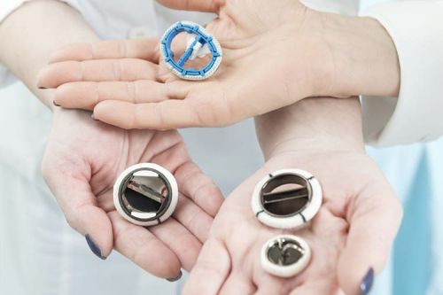
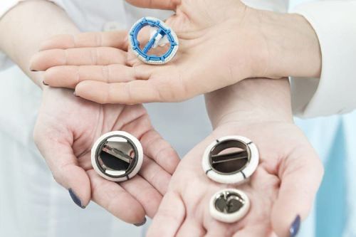
Today there are 3 types of valves used to replace damaged heart valves of patients, including: mechanical heart valves, biological heart valves and autologous valves. The specialist will be based on factors such as: Age; the patient's condition (degree of heart valve damage, degree of myocardial damage, clinical symptoms, underlying comorbidities,...); conditions for monitoring and treatment after valve replacement of the patient as well as economic conditions to advise which type of valve is suitable for each patient.
2.1. Mechanical Valves Mechanical heart valves are artificial valves made from long-life materials such as metal, carbon, ceramic and plastic.
The advantage of this valve is that the valve has a high durability (maybe over 20 years) if the patient adheres to the postoperative treatment as directed by the doctor. Therefore, mechanical valves are often recommended for young people to avoid having to change the valve many times. Another advantage is that mechanical valves are cheaper than biological valves. The disadvantage of using a mechanical valve is that the patient needs to take anticoagulants for life with the aim of preventing the risk of thrombosis on the valve (caused valve jamming, stroke complications, myocardial infarction, .. ). When taking anticoagulants (usually vitamin K antagonists), patients need to have their blood clotting index (INR) checked periodically to adjust the dose of the drug and the patient is also more prone to bleeding than people who do not take the drug (overdose). Anticoagulants can cause cerebral hemorrhage, gastrointestinal bleeding, bleeding in muscle and joint capsule, etc.). In addition, in pregnant women, the drug also increases the risk of teratogenicity, fetal malformation, especially in the first 3 months of pregnancy.
2.2. Biological valve This is a type of valve taken from the heart of an animal (heterozygous valve) that has been treated to remove components that cause transplant rejection for the patient and is partially repaired. They are placed on a metal or plastic support so that they can be placed on the body easily.
The advantage is that the patient does not have to take anticoagulants for life (usually only about 3 to 6 months after surgery).
The disadvantage is that because the biological valve is a natural valve tissue, it will gradually degenerate over time, affecting the function of the valve, causing re-stenosis or regurgitation. The average life of a biological heart valve lasts from 8 to 10 years and the degree of valve degeneration depends on age and pressure on the valve. The rate of biological valve degeneration is faster in young people, especially pregnant women, so biological valves are often indicated in patients over 60 years old.
Replacing the prosthetic valve in children needs to be done many times because the ring of the artificial valve is made of the natural valve, does not grow with the child and the leaflets become narrow as the child grows.
2.3. Replacing autologous heart valve by Ozaki method The technique of using autologous pericardium to regenerate aortic valve is a method invented by Professor Ozaki (Japanese) more than 10 years ago. The Ozaki method takes the patient's own pericardium to regenerate it into a heart valve for them, the pericardium is sewn directly into the natural valve ring and then acts like a natural valve, so blood flow through the valve is less obstructed. resistance than mechanical or biological valves. The technique of heart valve reconstruction with autologous materials has now been applied in more than 50 countries around the world and is considered a great step forward, bringing many advantages in heart valve replacement therapy such as: The disease does not require lifelong anticoagulation. Reduced risk of bleeding and infection when using prosthetic valves. For young people, the rate of not having to re-operate after 10 years for biological valves is 85%, while the rate of not having to have surgery again after 10 years if applying Ozaki technique to replace autologous valves accounts for 95 %. % - 98%. The time of the heart valve lasts almost the patient's life (because the heart valve is regenerated from a part of the patient's body, the body's tolerance will be better, limiting the rejection process, helping to limit the use of the heart valve). of the valve leaflet is elongated). The structure of autologous valves is similar to natural valves, so blood flow through the valve is less obstructed than mechanical or biological valves of animal origin. This method helps reduce treatment costs for patients because there is no need to buy artificial valves and repeat surgery many times. This method is especially suitable for patients who are women wishing to become pregnant, patients with small natural annulus, children in the developmental stage, patients with infections in the heart (endocarditis). conjunctivitis). Replacing autologous heart valves with the Ozaki method is an advanced technique, which should be performed by a qualified, highly qualified and well-trained specialist.
3. How is autologous valve replacement technique performed?
The pericardium is a membrane that surrounds the heart, before approaching the heart, the surgeon will cut a piece of pericardium about 7 x 8cm in size, then treat it with a special solution to the pericardium become more solid. After removing the damaged leaflet of the patient, the surgeon uses a special measuring instrument to measure the size between the two edges of the valve, then cuts the patient's pericardium according to the corresponding size based on the sample table. available. Then, in turn, each valve leaf is sutured to the patient's old valve position.
Ozaki method since its inception has helped patients with valve surgery reduce their worries. Starting in 2007, Dr. Shigeyuki Ozaki has implemented pericardial aortic valve reconstruction at Ohashi Hospital, Tokyo, Japan. In 2011, the author first announced the successful surgery results of 404 patients. Following that, in 2015, the author continued to publish successful results with a larger number of patients and many countries around the world have implemented this technique, of which Vietnam has applied in recent years. this method.
After learning from Prof. Ozaki and successfully applying the Ozaki method, some large hospitals in Vietnam have combined this minimally invasive technique with this Ozaki method. Patients with indications for aortic valve replacement are applied laparoscopic surgery to remove the pericardium with an incision only 6cm long, the reconstructed aortic valve acts like a natural valve, helping to treat effectively. aortic disease, patients reduce pain, do not have to open the entire sternum like the classic Ozaki method.
Hope the above information will help readers know more about autologous heart valve replacement method. It is hoped that this method will be more widely available so that more and more patients can benefit from this new technique.
Please dial HOTLINE for more information or register for an appointment HERE. Download MyVinmec app to make appointments faster and to manage your bookings easily.


