This is an automatically translated article.
Article by Doctor Pham Quoc Thanh - Department of Diagnostic Imaging - Vinmec Hai Phong International General Hospital.
To evaluate and detect brain lesions, cranial magnetic resonance imaging (MRI) is a modern, optimal, non-invasive imaging method. Through images obtained from cranial magnetic resonance imaging, the doctor will quickly diagnose and accurately detect abnormalities here, especially brain tumors, inflammation, brain vascular malformations...
1. What is brain magnetic resonance imaging?
The brain is a particularly important organ that coordinates the activities of all organs and parts in the human body, is the control center of the central nervous system, responsible for controlling behavior. The brain is protected by the outer covering of the skull, a hard skeleton that, although the skull protects the brain parenchyma from injury, interferes with the study of brain functions in both normal and diseased conditions.
Brain magnetic resonance imaging (MRI) is an advanced imaging technique that uses magnetic fields and radio waves to produce detailed images of the brain and brain stem, with the advantage of not using radiation. invasive compared with other imaging methods such as computed tomography (CLVT), X-ray, digital erasure scan (DSA)... should be commonly applied in the diagnosis of neurological diseases . With this technique, without the need for contrast injection, we can still obtain detailed and clear images of skull abnormalities, especially brain tumors, with 3-D localization that other methods cannot do.
A magnetic resonance imaging machine is a system consisting of a large magnet with a central tunnel. The patient when taking the picture will be placed on a table that is inserted into this tunnel. During the scan, radio waves hit the magnetic positions of H+ atoms in the body and send out a signal that is sent to an antenna and a computer. The computer performs calculations to produce black and white images of cross-sections of the skull. Images of the area being examined can be converted into 3D to make it easier to identify abnormalities in the brain.
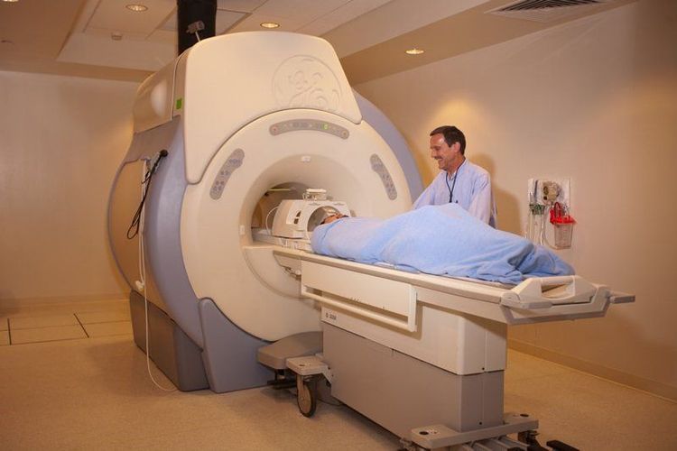
Chụp cộng hưởng từ não là phương pháp chẩn đoán hình ảnh hiện đại
Basically, cranial MRI is a non-invasive method, but the images give a high-resolution grayscale indicator, which helps to evaluate and detect lesions and abnormalities of the skull very early. Not only that, this method also evaluates brain function, sensory function as well as motor function, detects abnormal tumors, metabolic disorders...so it should be evaluated by medicine. very high.
2. What do brain magnetic resonance imaging results show?
Can detect many pathological conditions of the brain as well as abnormalities in brain structure such as tumor, cyst, hemorrhage, edema, inflammation, infection.. Observe the brain circuit without the need for plasma injection. Contrast.Cerebral vascular diseases: aneurysms, cerebral vascular malformations: arteriovenous malformations, venous malformations, cavernosal carotid artery shunts... Provide clear images of brain parenchymal components which can hardly be seen with conventional computed tomography (CLVT), ultrasound or X-ray alone. Because of that, cranial magnetic resonance imaging (MRI) helps diagnose brain stem and pituitary diseases extremely effectively. Plays a role in evaluating other problems such as: persistent headache, muscle weakness or paralysis, nervous system diseases. chronic menstruation; Clear, detailed images of brain tumors; Assessment of bleeding in the meninges, traumatic brain injury Detecting early stages of stroke. Evaluation of parts that are hidden by bone or difficult to see when taken with other methods.
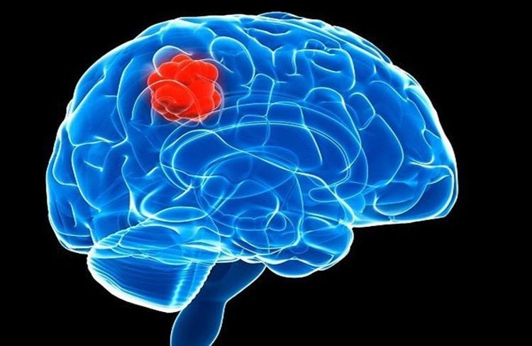
Chụp MRI giúp chẩn đoán các bệnh lý thân não hiệu quả
3. Benefits of MRI and combined with other imaging methods
Magnetic resonance imaging: The patient is not affected by radiation such as X-ray, CT scan or digital erasure scan (DSA), so it is not biologically affected, not radioactive.
High contrast soft tissue imaging, detailed anatomical description, more sensitive and specific abnormalities in the brain, areas covered by skull bones should evaluate the discrimination better than imaging CVT.
In emergency cases, especially in trauma such as traumatic brain injury, abdominal cavity, CT scan is an advantage because of the fast capture time, independent of the patient's movement, the patient has metal, Implants and especially calcified, gaseous lesions will be difficult to evaluate on magnetic resonance.
Vascular abnormalities such as cerebral aneurysm, cerebral vascular malformation, and embolism when detected early on MRI without contrast injection on cranial magnetic resonance imaging will be monitored for the progression of lesions, helping to treatment of aneurysms, cerebrovascular malformations, thrombectomy when combined with cerebral angiography, imaging and treatment of brain vessels digitally erasing the background and monitoring recurrence after treatment.
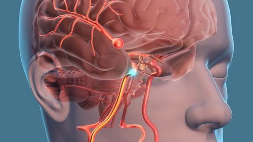
Chụp cộng hưởng từ phát hiện dị thường mạch máu não
4. When should cranial magnetic resonance imaging?
4.1. People with the following symptoms should have a cranial magnetic resonance imaging (MRI) scan of the brain. It is recommended to consult a specialist about brain MRI when having the following symptoms:
Dizziness for a long time does not improve. Headache persists in different degrees, migraine or whole head, dull pain or attacks... Memory is markedly impaired: poor thinking, decreased ability to concentrate, confusion, forgetfulness ...; Frequent insomnia, poor sleep, difficulty falling asleep, or waking up early without being able to go back to sleep... Hemiplegia, the person is so weak that it is difficult to grasp, difficulty in movement, sensory disturbances, or falling food One side... Visibly reduced vision or pain on one side. Distorted mouth, distorted face, difficulty speaking or hearing. Constipation, stiff neck, vomiting, convulsions, seizures.
4.2. Medical cases requiring cranial magnetic resonance imaging Some of the following cases require cranial magnetic resonance imaging to detect internal pathology:
Traumatic brain injury. Cranial nerve tumor, brain tumor. Cerebral infarction, cerebral hemorrhage, cerebrovascular accident. Meningitis, encephalitis. Cerebral vascular malformation, cerebrovascular disease, neuro-vascular conflict. Birth defects in the brain: brain defects, brain atrophy.... Multiple sclerosis, white matter degeneration. Follow-up after brain surgery.

Các bệnh lý bên trong não cần phải chụp cộng hưởng từ sọ để chẩn đoán
5. Magnetic resonance imaging (MRI) of the brain at Vinmec Hai Phong International General Hospital
Vinmec Hai Phong International General Hospital is currently one of the hospitals in the Vinmec International Hospital system equipped with modern machinery and equipment for general medical examination and treatment and magnetic resonance imaging (MRI). MRI) accurate, modern for diseases of the brain, cerebrovascular in particular.
Patients performing MRI at Vinmec Hai Phong International General Hospital will be clearly guided by the support staff and technical staff on the manipulations, scanning procedures, best safety issues, and guaranteed safety. for good diagnostic images without harming the patient.
Especially, Vinmec Hai Phong International General Hospital is the first unit in Hai Phong to put into use the new 3.0 Tesla Silent Magnetic Resonance Scanner from the US manufacturer GE Healthcare. The machine applies the safest, most accurate, non-invasive magnetic resonance imaging technology available today. Silent technology is very beneficial for patients who are young children, the elderly, patients with weak health or have just had surgery.
To register for examination and treatment at Vinmec Hai Phong International General Hospital, you can contact Hotline 0225 7309 888, or register online HERE.
Reference source: Medicalnewstoday.com; webmd.com


