This is an automatically translated article.
Pulmonary perfusion scintigraphy is an advanced nuclear scanning technique used to find blood clots that block normal blood flow in the lungs (pulmonary embolism).
1. Overview of pulmonary perfusion scintigraphy in pulmonary embolism
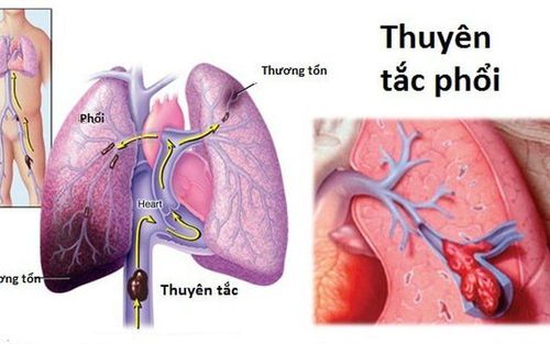
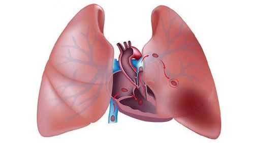
Scanning is a method of taking pictures of the bones or organs of the body by inserting a small amount of radioactive tracer into the body. A lung scan (also called a “perfusion lung scan” or a “VQ scan”) can help detect blood clots that are present in the lungs (pulmonary embolism).
There are 2 types of lung scans done alone or together:
Ventilation scan: During this scan, the patient is given a breath of air or a radioactive tracer to help document the areas. of the lungs lack air or hold too much air. Ventilation scans are usually performed first. Pulmonary perfusion scan: The patient will be injected with a small amount of radioactive tracer (Galium Citrate) into a vein in the upper arm. This substance travels through the bloodstream and into the lungs helping to show areas of the lung that are not receiving an adequate dose of blood. If the lungs are functioning properly and normally, the results of both lung ventilation and perfusion scans will match. Conversely, if the scan results do not match, there may be a blood clot inside the lung.

Xạ hình thông khí tưới máu phổi giúp phát hiện những cục máu đông tồn tại trong phổi
2. Indications and contraindications
A pulmonary perfusion scan is often done to:
Find a thrombus that is preventing normal blood flow to the lungs. Check and evaluate the flow of blood or air through the lungs. Evaluation of which part of the lung is working and which is damaged, is done before lung surgery to determine which parts of the lung need to be removed. Pulmonary ventilation and perfusion scintigraphy is contraindicated in patients:
Severe pulmonary circulatory failure, acute pneumonia. Severe pulmonary hypertension. Sensitive to proteins.
3. Pulmonary ventilation perfusion imaging procedure
3.1. Preparation Prior to the pulmonary ventilation and perfusion scan, the patient should tell the doctor if:
Are or may become pregnant. Breastfeeding. Because radioactive tracer can pass into breast milk and affect the baby. Lactating patients should discontinue breastfeeding for 1 or 2 days after this procedure.

Phụ nữ mang thai hoặc đang cho con bú cần thông báo với bác sĩ trước khi tiến hành xạ hình
Patients should talk to their doctor with questions about the pulmonary ventilation perfusion scan procedure, its risks, or the implications of the results.
Before the procedure, the patient needs to remove jewelry or may have to undress to easily capture the area to be examined.
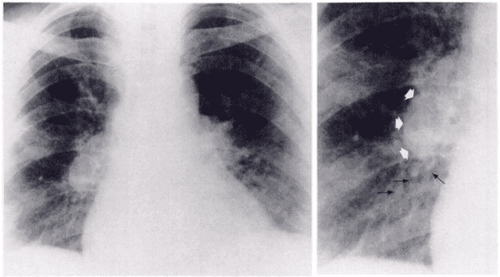
3.2 Pulmonary Perfusion Scanning Procedure Chest x-rays can be performed on the same day, before or after the pulmonary perfusion scan to depict abnormalities. The pulmonary perfusion scan is performed in the following steps:
Pulmonary ventilation scan:
To scan the lung ventilation, the patient will be placed 1 mask on the mouth and nose, or 1 nose clip combined with tube in the mouth to breathe. The patient is asked to take several deep, short breaths (about 10 seconds) to allow the camera to capture the radioactive tracer moving through the lungs. The camera moves and takes pictures from different positions. The patient needs to remain still during the scan for the best image quality. Once done, the radioactive gas will clear itself from the lungs when breathing. The lung ventilation scan is expected to take 15-30 minutes. Pulmonary perfusion scan:
The patient is asked to lie on his back, intravenously a small amount of radioactive tracer. After the injection, the camera captures an image of the tracer moving through the lungs. The camera is placed around the chest to get different angles. The patient needs to remain still during the scan to avoid image shake. The pulmonary perfusion scan is expected to take 15-30 minutes.
4. Notes after scanning the lung ventilation and perfusion scan
The results of the pulmonary perfusion scan may not be useful in the following situations:The patient is pregnant (because the amount of radiation from the scan can affect the fetus).

Quét xạ hình thông khí tưới máu phổi có thể không hữu ích nếu bệnh nhân có thai
Patient does not lie still during lung scan. The patient cannot breathe through a mask or tube. Patient has a medical condition involving the heart or lungs, such as chronic obstructive pulmonary disease (COPD), acute pulmonary edema, etc. If the pulmonary perfusion scintigraphy is not clear or helpful enough , your doctor may recommend another imaging test, such as a pulmonary angiogram, to help check blood flow to your lungs.
People with pulmonary embolism, in addition to medical treatment, should combine it with lifestyle changes and living habits. Even after successful treatment of pulmonary embolism, patients should also adopt these habits to prevent recurrence of the disease. Including:
Follow-up check-ups on schedule so that doctors can control the disease, promptly detect abnormal developments Take the right dose of medicine, according to the doctor's instructions. Do not arbitrarily stop smoking, do not arbitrarily buy drugs without the doctor's permission Limit lying down for too long Exercise regularly, regularly move Do not smoke, do not use stimulants Weight control If you are overweight, you need to lose weight if you are obese. Try to keep your toes higher than your hips when lying down or sitting. Do not wear clothes that are too difficult to prevent blood circulation. Currently, at Vinmec International General Hospital, fibrinolysis is being applied to emergency treatment of patients with pulmonary embolism. With modern imaging equipment, CT perfusion results are available within 7 minutes and performed routinely. The technique is performed by a team of qualified, experienced doctors and modern equipment and professional services.
Please dial HOTLINE for more information or register for an appointment HERE. Download MyVinmec app to make appointments faster and to manage your bookings easily.


