This is an automatically translated article.
Article written by Dr. Pham Quoc Thanh - Diagnostic Imaging Department - Vinmec Hai Phong International General Hospital
Digital Subtraction Angiography (DSA) is an X-ray angiography system with a combination of conventional Seldinger angiography with image processing techniques. by computer.
1. What is the DSA Structure System?
A DSA system usually consists of components such as an x-ray generator (lamp head), image acquisition unit, digital image processor and display unit...
The central part of the system That system is a digital image processing system. It will collect images from video camera and generate pulse control signal in time for both X-ray generator and image acquisition system in order to control the signal reception and transmission between the two systems. this.
The basic components of the DSA system:
Image Intensifier: The main purpose of using an intensifier is to reduce the patient dose and ensure the quality of the image obtained. Light Aperture Lens system Similar to the camera lens system, it is located at the output of the optical tube to change the amount of light entering the video camera for each dose. Specifically.

Hệ thống máy chụp mạch số hóa xóa nền (DSA)
Video Camera: is one of the key parts of digital X-ray system in general and DSA in particular. Its main function is to generate an analog electronic signal proportional to the amount of light received through the lens system (light aperture). Digital image processor: The digital image processor is the central part of the DSA system. It performs the main functions such as: Acquisition and digitization of video images. Store digital images in memory. Implement algorithms on archived images. Display the image on the monitor screen. Store images and data in hard disks or magnetic optical discs. The image processor usually includes a microprocessor or a computer control system to control and control the operation of the X-ray emission process, the data processing process.
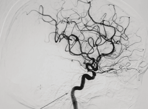
2. Principle of background erasing digital angiography machine
Before conducting digital erasure angiography (DSA), the doctors (technicians) will put a catheter into the blood vessels to examine by Seldinger method, then inject contrast agent through the catheter with effective Used to brighten blood vessels, this contrast is completely harmless and will be eliminated from the body in the patient's urine.
When X-rays are emitted, they pass through the patient's body and are picked up by the Image Intensifier. A lens is placed between the bulb and the video camera to limit the amount of light reaching the camera. Before performing the background removal algorithm, the camera captures the initial image without contrast injection. The contrast material is then injected into the blood vessel to be imaged through a catheter inserted into the femoral artery through the skin. Next is the process of acquiring an animated image when the contrast agent enters the body in a preset time unit. The image processing unit will take the image obtained when there is no contrast agent as the background image (mask image) and proceed to exclude the background image with the image obtained when the contrast is present, which are static anatomical structures like between two images.
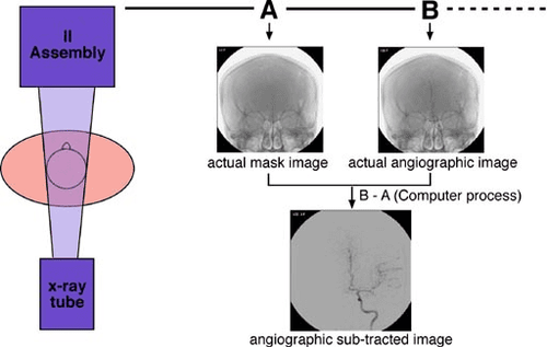
Nguyên lý của máy chụp mạch số hóa xóa nền
3. What do patients need to prepare when taking digital angiography?
Before taking DSA background digitized angiogram, the patient must be carefully prepared: blood test, chest X-ray, electrocardiogram recording and fasting.
DSA scanning technique is an invasive technique and has a risk of complications when taking, so it must be strictly specified and the patient understands the technical process and agrees to do the intervention procedure.
The patient is inserted a catheter into the lumen of the blood vessel from the femoral artery (or brachial artery, radial artery) to the artery to be evaluated for contrast injection. The patient needs to cooperate and lie still during the intervention.
After the scan, the patient will be bandaged in the groin area, immobilized, and immobilized at the puncture site for 8-10 hours.
4. Is background erasing digital angiography dangerous?
The DSA procedure is usually painless, and you may feel some discomfort when the injection is given at first. If you are too stressed, your doctor may give you a sedative, but this is not usually necessary.
Chụp mạch máu kỹ thuật số hóa xóa nền không gây đau đớn cho người bệnh
Some possible complications after performing DSA are classified into local and systemic complications, including:
Local complications (at the injection site):
Blood clot formation Injury nearby tissue Pseudoaneurysm Arteriovenous fistula Systemic complications:
Contrast allergy Thrombotic embolism Gas embolism Vessel dissection Decreased function, renal failure For detailed advice , please come directly to Vinmec health system or register online HERE.