This is an automatically translated article.
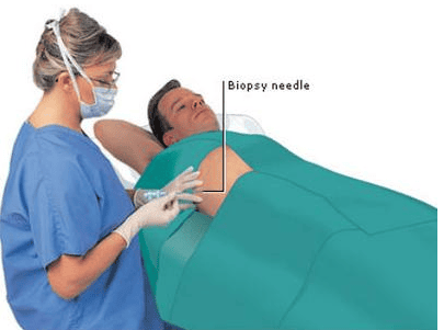
The article is professionally consulted by MSc, BS. Dang Manh Cuong - Doctor of Radiology - Department of Diagnostic Imaging - Vinmec Central Park International General Hospital.Liver biopsy is a commonly used procedure in the diagnosis and treatment of liver diseases. Normally, patients have percutaneous liver biopsies, in cases of contraindications to percutaneous biopsies, digital erasure of the background and liver biopsy through the internal jugular vein can be applied.
1. What is a digital background scan and liver biopsy through the internal jugular vein?
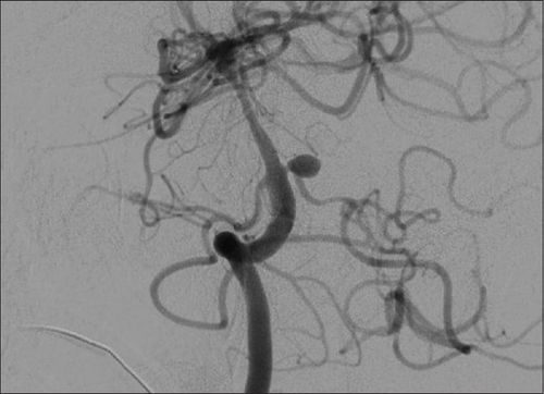
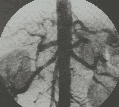
Liver parenchymal tissue is mainly obtained by percutaneous biopsy under ultrasound guidance or computed tomography. However, in some cases where a percutaneous biopsy is contraindicated, a liver biopsy can be performed via the internal jugular vein by using an instrument from the internal jugular vein, through the venous system. superior vena cava – inferior vena cava, enter the hepatic vein and puncture the hepatic vein wall to remove liver tissue.Digital Subtraction Angiography (DSA) is a new vascular imaging technique using X-rays. The purpose of this technique is to study blood vessels in the body and better see lesions and pathologies. blood vessels before vascular intervention is indicated. This is an imaging method that combines X-ray and digital processing using algorithms to remove the background on 2 images obtained before and after injecting contrast material into the patient's body.
Sinh thiết gan qua tĩnh mạch cảnh trong được áp dụng cho một số trường hợp chống chỉ định sinh thiết qua da.
2. In which cases are digital scans to erase the background and biopsies of the liver through the internal jugular vein?
This technique is indicated in the following cases:In cases where a biopsy of the liver parenchyma is required but a percutaneous biopsy is contraindicated, such as coagulopathy, thrombocytopenia or a lot of free intra-abdominal fluid. Preoperative assessment for patients preparing for liver transplantation Diffuse liver disease. Digital erasure of the background and liver biopsy through the internal jugular vein are contraindicated without the consent of the patient or the patient's family. Relative contraindications for pregnant women, severe coagulopathy

Phụ nữ mang thai chống chỉ định thực hiện
3. How are digital background scans and liver biopsies performed through the internal jugular vein?
Before the procedure, the patient is thoroughly explained about the procedure to coordinate with the doctor, the patient is also asked to fast before 4 hours. At the beginning of the procedure, if the patient is too excited to lie still, he or she may be given sedation. The patient requires general anesthesia. Intravascular access: Needle puncture into the internal jugular vein, usually performed under ultrasound guidance. Insertion of a catheter into the internal jugular vein Angiography: Insert the catheter into the superior hepatic vein, usually the right superior hepatic vein. Angiography of by regurgitation of contrast from the superior hepatic vein through the liver parenchyma into the vein. Liver biopsy: Insert the tube into the 9F vein into the internal jugular vein, possibly inserting the tip of the tube into the lumen into the orifice of the right superior hepatic vein. An angiogram of the liver to determine the site to be biopsied. Measure hepatic sinus pressure to assess cirrhosis. Proceed to cut the specimen through the tube into the vascular lumen. After the intervention, the patient will be monitored within 6 hours: hemodynamic status, pulse, blood pressure to prevent complications of intra-abdominal bleeding or perforation of hollow viscera due to the needle passing through the liver capsule.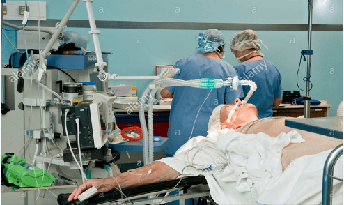
Trong quá trình làm thủ thuật, bệnh nhân cần gây mê toàn thân
4. Possible complications when performing the procedure
Cardiac arrhythmia caused by sliding catheter or tube into the lumen into the right atrium. Pericardial hemothorax caused by the needle slipping into the right atrium, causing right atrium perforation. Puncture of the biliary tree or gallbladder causes biliary bleeding. Puncture of the hepatic artery causes pseudoaneurysm or intra-abdominal bleeding. Perforation of the liver capsule. Digital erasure of the background and liver biopsy through the internal jugular vein are difficult imaging techniques, requiring specialized technicians, and this technique must be performed at medical facilities. has full facilities, modern machinery system, standard photography room. Therefore, in order to achieve accurate imaging results, patients need to choose reputable addresses with digital background erasers and modern and standard medical equipment to perform. timely treatment.Vinmec International General Hospital is a hospital with a full convergence of general and specialized doctors to perform, examine, operate, diagnose and treat diseases. In particular, at Vinmec, it continues to invest in a system of modern machinery to serve the examination and treatment of diseases.
Besides, Vinmec has also performed digital imaging techniques to erase the background and liver biopsies in many cases to promptly provide results and treatment directions for many customers.
Before taking a job at Vinmec Central Park International Hospital from December 2017, Doctor Dang Manh Cuong has over 18 years of experience in the field of ultrasound - diagnostic imaging in Transport Hospitals. Hai Phong, MRI Department of Nguyen Tri Phuong Hospital and Diagnostic Imaging Department of Becamex International Hospital.
To register for examination and treatment at Vinmec International General Hospital, you can contact Vinmec Health System nationwide, or register online HERE