This is an automatically translated article.
The article was professionally consulted by Specialist Doctor I Tran Cong Trinh - Radiologist - Radiology Department - Vinmec Central Park International General Hospital.
Coughing up blood is a medical emergency, often with a variety of causes. If not treated promptly, the patient can die from hemorrhagic shock or respiratory failure. Currently, digital angiography and bronchial embolization are minimally invasive and highly effective procedures in the treatment of hemoptysis.
1. What is background erasure and bronchial embolization?
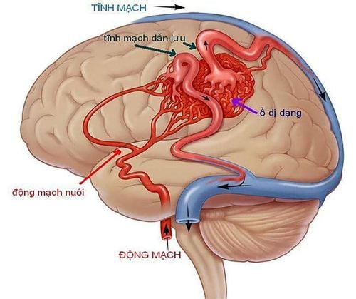
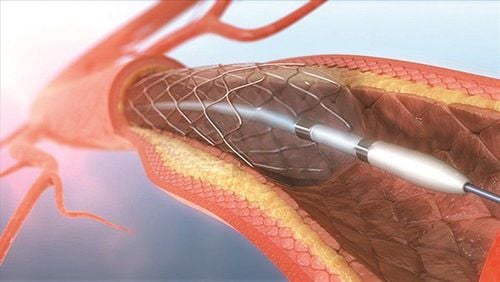
Pulmonary tuberculosis , lung tumors , pulmonary fungi , infections are common causes of hemoptysis . Traditional methods to stop bleeding and exit when coughing such as bronchoscopy to stop bleeding, surgery to clamp the bleeding or cut the damaged lung if necessary. However, with the development of current science, embolization method is an interventional endovascular treatment technique that is considered a less invasive method but brings high efficiency, safety, and few complications. Angiography and bronchial artery embolization were applied through the Digital Subtraction Angiography (DSA) system. The basic principle of the DSA imaging system is to use fluorescence light and X-ray angiography in the areas to be examined before the contrast medium has been injected and after the contrast material has been injected into the blood vessels to be examined. take a shot. Finally, to clarify the vascular system, the computer will erase the background image.Angiography and embolization of the bronchial artery is to visualize this artery on the screen by injecting contrast agent directly into the artery, entering from the femoral artery, and plugging the branches with specialized materials. The bronchial artery originates from the thoracic aorta from the anterior surface at the level of the D4-D5 thoracic vertebrae and divides into two right and left branches.

Chụp số hóa xóa nền và nút mạch phế quản có vai trò quan trọng trong việc điều trị ho ra máu
2. Indications and contraindications of bronchial artery embolization and angiography
2.1. Point
Cases of severe hemoptysis, small amount but prolonged hemoptysis have not had a radical surgical condition or no indication for surgery.2.2. Contraindications
Absolutely contraindicated in severe coagulopathy (prothrombin < 70%), platelets < 50 G/l. Relative contraindications for patients with severe organ failure, allergy to contrast agents.
Bệnh nhân rối loạn đông máu chống chỉ định thực hiện
3. How is the method of digital scan to erase the background and bronchial embolization performed?
Similar to other invasive procedures, before proceeding, the doctor will explain to the patient clearly about the technique as well as the steps to take so that the patient has a good coordination with the doctor and patient. They are also required to fast for 6 hours before the exercise, if they drink water, drink less than 50ml.3.1. The emotionless method
The patient will lie on his back on the table, place an intravenous line, pre-anesthetic or anesthetic if necessary (patient is not awake, young child...)3.2. Select the catheter technique and route of entry
According to the Seldinger method, the entrance can be from the femoral artery, axillary artery, brachial artery, carotid artery, and radial artery. Usually femoral artery, unless this route does not work, then choose other routes.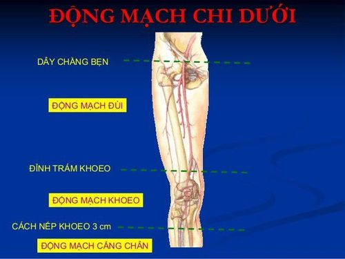
Động mạch đùi thường là đường vào của ống thông
3. 3. Performing the trick
The nurse disinfects the groin area on both sides (the way to the femoral artery). Spread stretch, cover the entire patient, leave the puncture site, the catheter inserted into the angiogram Local anesthesia at the site of insertion of the catheter into the lumen Needle puncture, place the kit in the lumen of the femoral artery. Insert the lead and angiographic catheter through the aortic initiator, find the origin of the bronchial artery, inject 3-5ml of the drug to confirm the bronchial artery with contrast agent through the syringe The machine can see the entire right and left bronchial artery system. If other branches are suspected to supply blood to the affected area, it is necessary to take full pictures of the branches (internal mammary artery, external thoracic artery, intercostal space, subphrenic) Obstruction of abnormally dilated arterial branches supplying blood to the affected area. the damaged area with specialized embolization materials (PVA beads, metal spirals, bio-glue, spongel..) Withdraw the catheter and the catheter into the lumen from the vessel after having taken and occluded the blood vessels as required, Apply pressure by hand for 15 minutes to stop bleeding, then apply pressure for 6 hours. After the procedure, the patient is asked to lie still on the bed, the leg on the side of the angiogram is stretched, immobile, the doctor will monitor the dorsal pulse, monitor bleeding, hematoma at the puncture site, and monitor the whole body. (pulse, blood pressure, patient's response)In summary, digitized scanning of the background and bronchial embolization is a modern and highly effective technique in the treatment of hemoptysis due to bronchial artery dilation. However, this is still an invasive procedure, so there is still a risk of complications during the procedure, so patients need to adhere to strict regulations before and after the procedure.
Digitization of the bronchial artery nodal background is an imaging technique that requires a high level of experience of the doctor and the perfect coordination of the patient. In order to achieve high diagnostic efficiency, patients need to choose reputable addresses that have digital scanning machines to erase the background and have modern and standard medical equipment from which to have timely treatment.
Vinmec International General Hospital is a hospital with a full convergence of general and specialized doctors to perform, examine, operate, diagnose and treat diseases. In particular, at Vinmec, the technique of digital imaging to remove the background is also performed to diagnose images, thereby offering treatment plans for many different diseases and providing optimal treatment results for customers.
Before taking a job at Vinmec Central Park International General Hospital, the position of Doctor of Radiology from September 2017, Doctor Tran Cong Trinh worked at Gia Dinh People's Hospital since 2007. -2017. In his role, Dr. Tran Cong Trinh has participated in guiding the teaching of students, residents, specialists and new doctors entering the department
For examination and treatment at International General Hospital Vinmec, please come directly to Vinmec Health System or register online HERE.