This is an automatically translated article.
The article is written by Master, Doctor Mai Vien Phuong - Gastroenterologist - Department of Medical Examination & Internal Medicine - Vinmec Central Park International General Hospital.Ultrasound is an imaging technique that uses ultrasound waves (high frequency sound waves) to build and reconstruct images of the internal structures of the body. Ultrasound is one of the most widely used diagnostic and therapeutic methods in medicine. The following article will clarify the role of ultrasound in the diagnosis and evaluation of bleeding ulcerative colitis.
1. Clinical manifestations of bleeding ulcerative colitis
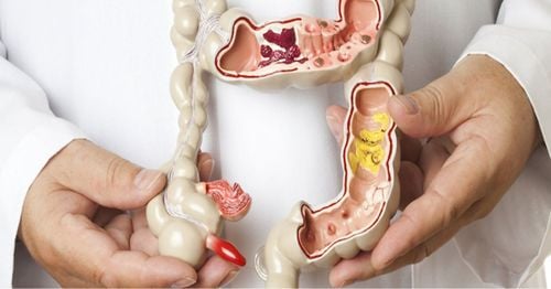
Hemorrhagic ulcerative colitis is characterized by diffuse and superficial inflammatory lesions of the colonic mucosa, beginning in the rectum and extending to other segments of the colon. The small intestine is usually not involved, although the terminal ileum may have superficial inflammatory lesions. Based on the extent of colonic lesions, ulcerative colitis can be classified into the following types: Proctitis (localized lesions in the rectum), sigmoid colitis - rectum or left colon. (spread to the splenic angle) or diffuse/global colitis. The extent of the lesion is not only related to severity but also affects wages and treatment options. Symptoms and course of disease are related to the extent and severity of inflammatory lesions. Symptoms are often insidious, although the disease may develop acutely after an episode of infectious colitis or travel diarrhea. Proctitis causing bloody stools is the most common symptom of patients. The degree of bloody stools can be severe, profuse or moderate, with bloody and mucusy stools. The patient may have frequent urge to defecate and strain or increase the frequency of defecation. The more widespread the colonic lesion, the more severe the diarrhea, while if the patient is simply proctitis, the symptoms may alternate between episodes of constipation with episodes of bloody stools.Abdominal pain before defecation or a feeling of abdominal distension will occur when the disease is advanced. In advanced or fulminant cases, the patient may present with systemic symptoms such as night sweats, fever, vomiting, nausea, weight loss accompanied by diarrhea. In addition, may experience extra-gastrointestinal symptoms such as in the eyes, skin, joints, liver.

Ulcerative colitis is diagnosed when the inflammatory lesion extends as far as the transverse colon or the right colon. Patients often present with diarrhea, bloody stools, painful defecation, abdominal fullness, abdominal cramps or localized pain. In addition, patients in this group often have weight loss, systemic symptoms, extra-gastrointestinal symptoms, and anemia. Toxic megacolon is the most severe complication of hemorrhagic ulcerative colitis when the inflammatory lesion spreads from the superficial mucosa to the submucosal and muscular layers. This complication often occurs in patients with disseminated colitis or severe colitis. Clinical manifestations include fever, exhaustion, severe abdominal cramps, abdominal distention, and local or total abdominal pain.
2. The role of ultrasound in the diagnosis and evaluation of bleeding ulcerative colitis
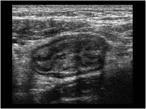
Hemorrhagic ulcerative colitis is a disease in which inflammatory lesions are mainly in the mucosa and are common in the rectum, so it is difficult to use imaging methods to evaluate. However, ultrasound is considered a reliable diagnostic tool to help assess the extent and activity of the disease. In particular, in cases where colonoscopy is contraindicated or cannot cover the entire colon.First, a Convex probe with a frequency of 3.5 - 5 MHz can be used for general evaluation. Then switch to the linear probe with frequency 4 - 13 MHz for detailed examination of the layers of the intestinal wall. It is possible to go from the epigastrium down or from the left iliac fossa (where the sigmoid colon is located) and then examine the colon, terminal ileum, appendix, small intestine, and up to the stomach. If the patient has pain localized to one site, a thorough examination is warranted. Assessment of damage:

Mucosal layer is usually an external hypoechoic line that is still preserved with regular borders and borders. Unlike in Crohn's, ulcerative lesions in bleeding ulcerative colitis are often superficial. Therefore, undetectable by ultrasonography and in mild to moderate stage, the inner layers of the intestinal wall are still evident. However, in the advanced stage of the disease, the intestinal wall will be clearly thickened, hypoechoic and the boundary between the inner layers of the intestinal wall is no longer soft, can see small hyperechoic dots representing the Ulcers eat into the submucosa. Especially, if the patient has toxic megacolon, the bowel wall in the left colon will be much thickened, hypoechoic along with dilatation of the transverse colon (>6cm), thin transverse colon wall. (< 2mm) and there was increased fluid in the bowel loops and dilatation of the ileal loops.

Please dial HOTLINE for more information or register for an appointment HERE. Download MyVinmec app to make appointments faster and to manage your bookings easily.


