This is an automatically translated article.
The article is professionally consulted by MSc, BS. Dang Manh Cuong - Radiologist - Radiology Department - Vinmec Central Park International General Hospital. The doctor has over 18 years of experience in the field of ultrasound - diagnostic imaging.1. What is background digitized visceral angiography?
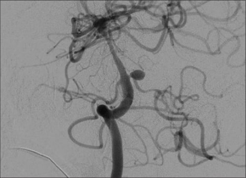
Background digitized venography is a modern imaging method, performed by injecting contrast material (iodine) through the arteries or veins, then capturing a raw image of the system. visceral veins. These images will be processed by a mechanical system, deleting irrelevant images, and clarifying the lesions (if any) of the visceral venous system.2. Indications and contraindications of digital splanchnic venography
2.1 Indications Diseases of malformed visceral venous system such as venous hemangioma, pseudoaneurysm of visceral vein...; Other visceral vein diseases such as venous stenosis; Scan to check the results after surgery to treat diseases of the venous system; Photograph of the visceral vein system digitally erases the background to serve the process of electro-optical intervention. 2.2 Contraindications The current method of digitalized visceral venous system removal has no absolute contraindications. Some relative contraindications of this method include:Patients with coagulopathy; Patients with renal failure, unable to use contrast agents; Allergic disease to contrast agents ; Pregnant women.

3. The person performing the DSA imaging technique of the visceral venous system
Specialist; Auxiliary physician; Electro-optical technician; Nursing; Doctor, anesthesiologist (if patient cannot cooperate).4. Means and drugs to prepare before taking a DSA of the visceral system
4.1 Machines and vehicles Digital background removal angiography (DSA); Dedicated electric pump; Film, film printer, image storage system; Lead shirt, apron to shield X-rays 4.2 Medicines to be prepared Local anesthetic; Pre-anesthesia and general anesthetic (if the patient has an indication for general anesthesia); Anticoagulants; Drugs that neutralize anticoagulants; Water-soluble iodinated contrast agent; Antiseptic solution for skin and mucous membranes 4.3 Prepare the patient before performing a DSA scan The patient is thoroughly explained about the procedure and procedure for a digital visceral angiogram to remove the background in order to cooperate with the doctor and the doctors. technicians. The patient or adoptive family member must write a written consent to the procedure before performing the procedure. Patients need to fast, fast for 6 hours before or can drink no more than 50ml of water. After entering the intervention room, the patient will lie in the supine position, and the technicians will install the machine to monitor vital signs such as breathing rate, pulse, blood pressure, electrocardiogram, etc. arterial blood gases. In case the patient is uncooperative or aggressive, a sedative will be prescribed. Disinfect the skin surface with an antiseptic solution and cover with a sterile towel with holes.
5. The process of digitizing the visceral vein system to remove the background
5.1 Anesthesia method The patient lies in the supine position on the DSA table, the technician places the line, usually using 0.9% physiological saline solution. In most cases, only local anesthesia is needed. However, with exceptions where pre-anesthesia is required, such as: children under 5 years of age, who do not have a sense of cooperation with doctors, or patients who are too excited or scared, etc., it is necessary to conduct general anesthesia. body throughout the procedure. 5.2 Selection of the catheter inlet The doctor uses the Seldinger technique to insert the catheter into the body. The access to the catheter is usually through large arteries such as the femoral artery, axillary artery, brachial artery, or radial artery. In most cases, it will be inserted through the femoral artery because this is a large artery, easy to insert the catheter. 5.3 Visceral vein angiography Sterilize and anesthetize the selected site to insert the catheter into the body; Insert the catheter and insert the tube through the selected path; Insert the catheter into the superior mesenteric artery system of the visceral artery system; Inject the contrast agent at a dose of 20-30ml, with a slow speed of 4-5ml/sec; Perform angiography in both arterial and venous phases to study abnormal images of the visceral venous system including the mesenteric and portal venous systems. 5.4 End of venography Withdraw the catheter from the patient's body; Apply pressure at the catheter puncture site by hand directly for 15 minutes, then apply pressure to fix it with an instrument for 6 hours or use a device to close the lumen; Advise and interpret results to patients.6. Complications of the visceral venous system scan
6.1 Complications during the procedure Arterial wall tear causing bleeding or dissection of the artery wall at the catheter insertion site. Treatment is by stopping the procedure immediately, applying local compression and monitoring closely. Continuation of imaging through contralateral arterial puncture; Catheter break and stay in the lumen: Use special tools to remove through endovascular intervention or surgery; Allergy to contrast agent.
A system of modern and advanced medical equipment, possessing many of the best machines in the world, helping to detect many difficult and dangerous diseases in a short time, supporting the diagnosis and treatment of doctors the most effective. The hospital space is designed according to 5-star hotel standards, giving patients comfort, friendliness and peace of mind.
Please dial HOTLINE for more information or register for an appointment HERE. Download MyVinmec app to make appointments faster and to manage your bookings easily.


