This is an automatically translated article.
Posted by Master, Doctor Mai Vien Phuong - Department of Examination & Internal Medicine - Vinmec Central Park International General Hospital
Hemorrhagic ulcerative colitis affects almost any age group from 15 to 35 years old. In order to examine and provide good treatments, you need to perform diagnostic tests as ordered by your doctor.
1. What is ulcerative colitis?
Ulcerative colitis (UC) is a type of inflammatory bowel disease (IBD). IBD includes a group of diseases that affect the gastrointestinal tract.
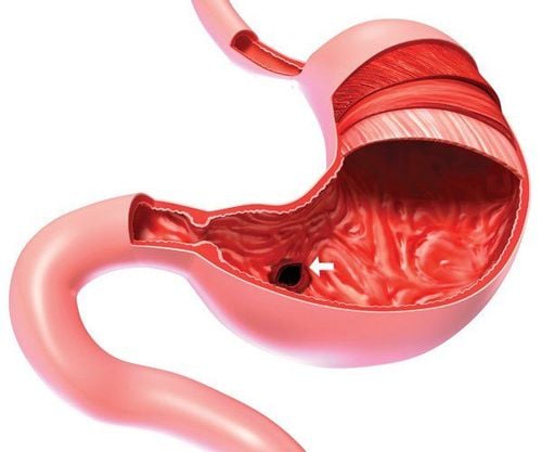
UC occurs when the lining of the large intestine (also called the colon), the rectum, or both becomes inflamed.
This inflammation creates small sores called ulcers on the lining of the colon. It usually begins in the rectum and spreads upward. The inflammation causes the intestines to move quickly and empty the intestines frequently. When the cells on the surface of your gut lining die, sores form. The sores may bleed, discharge mucus and pus.
While this disease affects people of all ages, most people are diagnosed between the ages of 15 and 35. After age 50, there is a small increase in men's diagnoses.
To learn more about the pathogenesis and clinical manifestations of bleeding ulcerative colitis, refer to the following article.
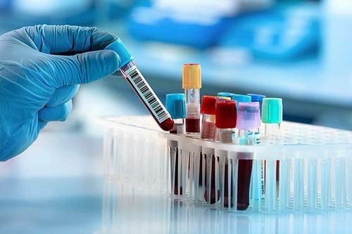
2. Subclinical used to diagnose bleeding ulcerative colitis
2.1. Test
Initial tests for patients with ulcerative colitis include complete blood count, electrolytes, liver and kidney function tests, iron levels, vitamin D, C-reactive protein (CRP), and calprotectin. in feces. Immunological and microbiological tests to rule out C. difficile should also be performed.2.1.1. Inflammatory indices
In patients with mild or moderate bleeding ulcerative colitis, inflammatory markers may remain within the normal range. In the results, a complete blood count may show thrombocytosis due to a chronic inflammatory response or to varying degrees of anemia. If leukocytosis is found, attention should be paid to infectious complications. In hemorrhagic ulcerative colitis, except for the group of patients with only localized rectal lesions, the CRP index was correlated with clinical severity. Elevated CRP is also correlated with increased erythrocyte sedimentation rate, anemia, and hypoalbuminemia. This is considered a prognostic marker in severe cases, with acute development. CRP above 10 mg/l after 1 year in patients with pancreatitis predicts an increased surgical risk. However, both CRP and erythrocyte sedimentation rate are not specific enough to differentiate bleeding ulcerative colitis from infectious or other causes of colitis.2.1.2. Fecal calprotectin test
A feature of chronic inflammatory bowel diseases including hemorrhagic ulcerative colitis is the infiltration of neutrophils into the glandular cleft of the intestinal epithelium and the epithelial lining. Therefore, there will be components of white blood cells in the stool. If there is no presence of leukocytes in the stool, it is possible to rule out chronic inflammatory bowel disease and follow other diagnostic directions.
In addition to the direct test for leukocytes in stool with methylene blue, there are now a number of stool tests that help detect leukocytes in the stool and are used to assess the severity of inflammatory bowel disease in the upper gastrointestinal tract. clinical. Calprotectin, derived from granulocytes, is a calcium-binding protein used as a diagnostic marker for cases of diarrhea caused by inflammatory bowel disease. In addition, this index also partly reflects the "inflammation level" of the colon, so it can be applied as a non-invasive method in the treatment process. Similarly, lactoferrin, a protein from neutrophils, has been shown to help differentiate between inflammatory bowel disease and irritable bowel syndrome.
Xét nghiệm Calprotectin trong phân giúp đánh giá mức độ nặng của bệnh lý viêm ruột lâm sàng
2.1.3. Microbiological test
As recommended by ECCO in 2017, microbiological tests should be done to rule out C.dificle and Cytomegalovirus (CMV) every outbreak. C. difficile infection is now becoming a serious health problem due to its high mortality rate. Therefore screening to exclude C. difficile should be performed if the patient is resistant to treatment or has a flare-up. CMV reactivation may occur in patients with bleeding ulcerative colitis receiving immunosuppressive agents. Although CMV reactivation is not the cause of sudden outbreaks, new CMV infection can make disease worse or become resistant to treatment. Therefore, patients on immunosuppressive therapy who experience flare-ups of symptoms should be tested for and ruled out for CMV infection.
Histopathology or immunohistochemical staining is more optimal than using PCR in blood.
2.1.4. Biomarkers
The two most commonly used biomarkers are anti-neutrophil cytoplasmic antibodies (DANCAS) and anti-Saccharomyces cerevisiae (anti-Saccharomyces cerevisiaeantibodies (ASCAs) antibodies). According to studies, the detection rate of pANCAS in patients with bleeding ulcerative colitis is up to 65% while the rate of this antibody positive in Crohn's patients is less than 10%. In contrast, ASCA is more specific for small bowel lesions due to Crohn's with a positive rate in 40-60% of cases. However, the positive rate of ASCA in other small bowel lesions such as Celiac is also quite high (40 - 60%).
Besides helping to differentiate between inflammatory bowel disease and other intestinal diseases, these biomarkers are also prognostic tools during treatment. High levels of pANCA are correlated with the risk of colostomy and ileostomy after total colectomy. Several other biomarkers are also being studied in IBD patients, such as antibodies to the outer membrane E.coli cell wall or J2 peptide which is an RNA sequence derived from E.coli. related to Pseudomonas fluorescens. Both of these markers have been shown to be more associated with Crohn's than hemorrhagic colitis and are not very effective in differentiating types of inflammatory bowel disease. The application of biomarkers in differentiating between ulcerative colitis and Crohn's is still difficult, especially in patients with atypical colitis. Using positive PANCA alone does not help distinguish between these two diseases, but adding ASCA can increase the rate of accurate diagnosis. Patients with pANCA+ASCA- are more common in ulcerative colitis with a sensitivity of 44-58% and a specificity of 81-98% while pANCA-ASCA+ is more common in Crohn's with small bowel involvement. with a sensitivity of 30 - 64% and a specificity of 92 - 97%. In the future, an additional combination of Ompc and 12 peptides is expected to help differentiate between bleeding ulcerative colitis and Crohn's.
2.2 Gastrointestinal endoscopy
2.2.1 Colonoscopy
Indications The 2017 ECCO Guidelines recommend colonoscopy with examination of lesions in the ileum, as an exploratory method to confirm the diagnosis of inflammatory bowel diseases.
In hemorrhagic ulcerative colitis, endoscopic lesions are initially in the rectal area close to the anal canal, then gradually spread upward with a continuous, concentric nature. The boundary between the lesion and the normal mucosa is clear. Occasionally, a localized cecum or no rectal lesion may be present, necessitating an assessment of the small bowel lesion. As in Crohn's, to accurately assess the lesion for hemorrhagic ulcerative colitis, multiple biopsies from the terminal ileum and five segments of the colon (upper, transverse, descending colon) are required. sigmoid colon, rectum). Ideally, each biopsies should include two sections of the normal mucosa and the lesion. When endoscopic and histopathological results are not clear, colonoscopy with biopsies can be performed in combination with other investigations such as upper gastrointestinal endoscopy if the patient is symptomatic, capsule endoscopy. or colonoscopy. For cases where the disease recurs, is resistant to treatment, appears with new symptoms or considers surgery, it is necessary to re-evaluate the extent of the damage endoscopically.
Assessment of lesion location In the Montreal classification, based on the location of the lesion, it is divided into the following categories: rectal lesions, left colon lesions and extensive lesions. Over time, the distribution of lesions may change, often tending to spread from the rectum to the upper colon. One study noted that 28% of cases after 10 years will have a more extensive lesion progression than at baseline.
Upper gastrointestinal endoscopy The upper gastrointestinal tract lesions in ulcerative colitis with bleeding are uncommon, so the indication for upper gastrointestinal endoscopy is made when the patient is symptomatic. Several reports have reported upper gastrointestinal tract lesions in patients with hemorrhagic ulcerative colitis, but the criteria for diagnosing upper gastrointestinal lesions are associated with bleeding ulcerative colitis until now. has not yet been agreed.

Nội soi đại tràng là phương pháp thăm dò giúp chẩn đoán bệnh lý viêm ruột
2.3. Other imaging methods
2.3.1.Ultrasound
Hemorrhagic ulcerative colitis is a disease in which inflammatory lesions are mainly in the mucosa and are common in the rectum, so using imaging methods to evaluate is difficult.
However, ultrasonography is considered as a reliable diagnostic tool to help assess the extent and activity of the disease, especially in cases where endoscopy is contraindicated or cannot cover all Colon. The Convex transducer with a frequency of 3.5 - 5 MHz can be used first for general evaluation and then switched to a linear probe with a frequency of 4 - 13 MHz for detailed examination of the layers of the intestinal wall. It is possible to go down from the epigastrium or from the left iliac fossa (where the sigmoid colon is located) and then examine the colon, terminal ileum, appendix, small intestine, and up to the stomach. If the patient has localized pain, a closer examination is necessary.
2.3.2. Take CT/CHT
Up to now, in ECCO guidelines as well as consensus between ECCO and ESGAR, there are no specific recommendations on indications and value of abdominal CT scan in assessing the extent and disease activity of patients. bleeding ulcerative colitis. Colonic enema technique can be used in CT scan with drug injection so that the arrowhead - thickening of the Sigma colon wall, small arrow - the lesion can be seen as the enhancement of the intestinal wall and the open tissue around the colon. colon more clearly. However, this approach is not indicated when the patient is suspected of having toxic dilated colonic aneurysm, perforation, or peritonitis.
Vinmec International General Hospital is one of the hospitals that not only ensures professional quality with a team of doctors, modern equipment and technology, but also stands out for its examination, consulting and service services. comprehensive and professional medical treatment; civilized, polite, safe and sterile medical examination and treatment space. Customers when choosing to perform tests here can be completely assured of the accuracy of test results.
Please dial HOTLINE for more information or register for an appointment HERE. Download MyVinmec app to make appointments faster and to manage your bookings easily.
Articles refer to sources: NagreF, Gionchetti PR, Eliakim R.(2017). Third European Evidence-based consensus on Diagnosis and Management of Ulcerative Colitis, De Dombal F.T. (1968), Ulcerative colitis: definition, historical background, aetiology, diagnosis, naturel history and local complications, Postgrad Med, Basler RW and Dubin HV (1976). Ulcerative colitis and the skin.