This is an automatically translated article.
Hemorrhagic stroke is a spontaneous bleeding from the cerebral arteriovenous system into the brain tissue or the ventricular system or the subarachnoid space. Craniotomy to remove hematoma is a method of treating stroke with cerebral parenchymal bleeding.
1. Hemorrhagic stroke
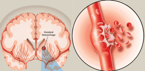
Hemorrhagic stroke is a medical condition caused by a hematoma caused by a ruptured blood vessel in the brain. A hemorrhagic stroke causes an area of the brain or areas of the brain that is deprived of blood supply, or that blood supply is greatly reduced due to narrowing of the arteries in the brain or a complete blockage.
Hemorrhagic stroke is divided into 2 types, depending on the location of bleeding
Intracerebral hemorrhage: A condition that occurs when a blood vessel in the brain bursts. Risk factors include high blood pressure, substance use, and alcohol consumption. In addition, it can also be caused by a leaky artery defect. Subarachnoid hemorrhage: A condition that occurs when a blood vessel on the surface of the brain bursts. Blood partially fills the space between the brain and skull, mixing with cerebrospinal fluid. When blood flows into the cerebrospinal fluid, it increases pressure on the brain and causes a severe headache.

Đột quỵ xuất huyết não do vỡ mạch máu trong sọ gây ra
2. Surgery to reduce pressure in hemorrhagic stroke
The majority of strokes with intracerebral hemorrhage are usually treated medically, the rate is about 90%, a few remaining cases require surgical intervention. In particular, life-saving measures for patients with increased intracranial pressure are also very effective and popular measures in the world today. Surgical treatment of cerebral hemorrhage and cerebral infarction in the following pathological cases:2.1 Hemorrhagic diathesis The following surgical indication criteria:
Glasgow score <9 points, consciousness gradually decreasing Volume hematoma >30g Widespread cerebral edema around the hematoma On CT or MRI Scan midline deviation >1cm In addition, exclusion criteria when indicated for surgery:
Blood pressure on maintenance vasopressor or mechanical ventilation Treatment results Surgical treatment has improved well Consciousness is good (Glasgow >= 13-15 points) Very deep coma <6 points Two dilated pupils with loss of light reflex 2.2 Thrombocytopenia Bleeding when indicated Surgery is also based on criteria such as fascia bleeding
2.3 Lobar bleeding The surgical indications for lobar bleeding include:
Glasgow score < 9 points Hematoma volume > 30ml Edema brain spread On CT or MRI Scan, the midline is more than 5mm displaced. Language and motor functions are getting worse

Phẫu thuật giảm áp là phương pháp can thiệp ngoại khoa hữu hiệu và phổ biến
2.4 pontic bleeding pontic bleeding is mainly medical treatment
2.5 intraventricular bleeding Intraventricular bleeding can drain the ventricles to reduce intracranial pressure
2.6 cerebellar bleeding Cerebellar hemorrhage needs to be surgically intervened as soon as possible, in order to prevent complications such as cerebellar amygdala, dilated ventricles causing increased intracranial pressure. Indication criteria for surgery
Consciousness <10 points, Glasgow gradually decrease Severe headache, agitation Hematoma volume >20g On imaging CT Scan, MRI Scan: displacement of IV ventricle, with dilated ventricle Lateral and III patients with posterior fossa hematoma usually return to normal life after timely surgical intervention
2.7 Infarct Infarct Criteria for surgical indications for cerebral infarction
Consciousness gradually decreases glasgow < 9 points Infarct area Heavy blood, nearly half of cerebral hemisphere On CT Scan and MRI Scan, midline deviation >1cm In summary, decompression surgery in hemorrhagic stroke is a commonly used surgical method today. Stroke has a high risk of death, so when you see abnormal signs such as distorted mouth, hemiplegia, ..., it is necessary to immediately go to a medical facility for examination and timely intervention.
Currently, Magnetic Resonance Imaging - MRI/MRA is considered a "golden" tool for brain stroke screening. MRI is used to check the condition of most organs in the body, especially valuable in detailed imaging of the brain or spinal nerves. Due to the good contrast and resolution, MRI images allow to detect abnormalities hidden behind bone layers that are difficult to recognize with other imaging methods. MRI can give more accurate results than X-ray techniques (except for DSA angiography) in diagnosing brain diseases, cardiovascular diseases, strokes,... Moreover, the process MRI scans do not cause side effects like X-rays or computed tomography (CT) scans.\
Vinmec International General Hospital currently owns a 3.0 Tesla MRI system equipped with state-of-the-art equipment by GE. Healthcare (USA) with high image quality, allows comprehensive assessment, does not miss the injury but reduces the time taken to take pictures. Silent technology helps to reduce noise, create comfort and reduce stress for the client during the shooting process, resulting in better image quality and shorter imaging time. With the state-of-the-art MRI system With the application of modern methods of cerebral vascular intervention, a team of experienced and well-trained neurologists and radiologists, Vinmec is a prestigious address for stroke risk screening and screening. reliable goods.
In the past time; Vinmec has successfully treated many cases of stroke in a timely manner, leaving no sequelae: saving the life of a patient suffering from 2 consecutive strokes; Responding to foreign female tourists to escape the "death door" of a stroke;...
Please dial HOTLINE for more information or register for an appointment HERE. Download MyVinmec app to make appointments faster and to manage your bookings easily.


