This is an automatically translated article.
Article by Specialist Doctor I Tran Ngoc Thuy Hang - Resuscitation - Emergency Doctor - Emergency Resuscitation Department - Vinmec Central Park International General HospitalBrain damage often leaves dangerous complications and seriously affects health. In some cases not treated promptly has to increase the risk of death. The management and treatment of brain injuries should be done carefully at reputable medical facilities.
1. Traumatic brain damage
Approximately 25% of patients with traumatic brain injury require urgent surgery for a subdural or epidural hematoma to resolve brain compression. Neurosurgery consultation should be considered early. Since 20% of patients with severe traumatic brain injury will have concomitant cervical spine injury, it is important to immobilize the cervical spine until appropriate assessment can be made.
Penetrating and non-penetrating injuries are often associated with cerebral edema formation, cerebral contusion, or intraparenchymal hemorrhage. Because the skull cannot expand to accommodate the increased intracranial volume and the compensatory space in the subarachnoid space of the spinal canal is very limited, the ICP (intracranial pressure) often becomes elevated. Monitoring and treatment of elevated ICP is considered important in such patients.
2. General principles in the treatment of traumatic brain injury
Be sure to perform resuscitation according to ABCs (airway, respiration, circulation) Avoid hypotension and maintain systolic blood pressure > 90 mm Hg. Avoid hypoxemia (PaO2 <60 mm Hg or SpO2 oxygen saturation < 90%) Maintain head and trunk axis alignment to avoid jugular venous pressure Keep head of bed at 30° to 45° unless patient low blood pressure. Elevating the head promotes venous drainage and cerebrospinal fluid to the canal. Maintain PaCO2 at 35 to 40 mm Hg. Prophylactic hyperventilation is not recommended. Hyperventilation is recommended as a temporary measure to reduce elevated intracranial pressure. Cerebral blood flow usually decreases during the first 24 hours after head injury and hyperventilation should be avoided during this period to prevent further hypoperfusion. Use isotonic saline as maintenance solution, do not use hypotonic saline. Actively treat fever to maintain normal body temperature. Control harmful agitation with sedation if necessary. Use drugs with a short half-life to facilitate continuous and accurate neurological assessment. Maintain normal electrolyte homeostasis and treat hyper/hypoglycemia. Evaluation and treatment of coagulation defects. Provide nutrition to achieve adequate calorie replacement as tolerated. Prophylactic anticonvulsants are appropriate during the first week after traumatic brain injury. Give mannitol (0.25-1 g/kg IV bolus) or hypertonic saline (eg, 5-10 mL/kg 3% NaCl as quickly as possible) when signs of hernia or neurological impairment do not appear. due to other factors. Expert advice should be sought when considering the application of measures to increase osmotic pressure. Avoid steroids: These agents are contraindicated in patients with head trauma. Initiate appropriate intracranial pressure monitoring: For patients with a Glasgow Coma scale of 3 to 8 after resuscitation or a score of 9 to 12 with abnormal computed tomography results. For patients with normal computed tomography but at least two of the following: Age > 40, systolic blood pressure <90 mm Hg, unilateral or bilateral decortical or extensor position Maintain Lowest perfusion pressure compatible with adequate cerebral blood flow. Target cerebral perfusion pressures range from 50 to 70 mm Hg. Although patients with intact autoregulatory mechanisms can tolerate higher values. Ideally, pressures provide adequate oxygen and cerebral perfusion while intracranial pressure is maintained <20 mm Hg.
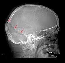
Hình ảnh chấn thương sọ não
3. Intracerebral hemorrhage
Patients with cerebral hemorrhage often have a history of hypertension. Blood pressure control is controversial in these cases. Elevated blood pressure may contribute to bleeding and the formation of cerebral edema but may also preserve local CPP (perfusion pressure).
Recent studies show that reducing systolic blood pressure to 140 mm Hg can improve outcomes. If elevated ICP is suspected, the patient should consult a specialist for assistance in blood pressure management.
Preferred agents include labetalol and nicardipine. The use of vasodilators is controversial, but drugs that cause significant intracranial vasodilation (eg, nitroprusside, nitroglycerin) should be avoided.
An increase in the volume of a hematoma is common, especially in patients taking anticoagulants or those with liver disease or thrombocytopenia. In some cases, surgery is considered to remove the hematoma, especially in children. When the patient clinically deteriorates with major lobar hemorrhage or when the bleeding is associated with a lesion that is surgically treatable, such as an aneurysm, arteriovenous malformation, or cavernous hemangioma. Deep basal ganglia has not been approved for routine surgical treatment.
4. Subarachnoid hemorrhage
Treatment of subarachnoid hemorrhage:
Make sure to perform resuscitation according to ABCs (airway, breathing, circulation)
Control blood pressure early, before definitive surgical treatment. Re-bleeding is an early complication until the aneurysm is clamped or rolled. Many intravenous drugs are useful. Labetalol and nicardipine have been advocated. Nitroprusside should be avoided because it tends to cause cerebral vasodilation. The effect of nimodipine on blood pressure should be considered when used in combination with other antihypertensive agents.
The patient started taking nimodipine, 60 mg, every 4 hours. (Some countries have intravenous preparations.), hypotension should be avoided.
Maintenance of intravascular volume: Some views favor a slight increase in intravascular volume. Significant heart damage can occur as a result of increased catecholamine levels. Therefore, careful attention should be paid to cardiac arrhythmias and cardiac function.
Avoid hyponatremia which is quite common. Isotonic saline should be used as the main intravenous fluid. Hyponatremia is more often a reflection of brain salt loss than SIADH (syndrome of inappropriate antidiuretic hormone). Cerebral salt loss should not be treated with volume restriction used for SIADH. Both groups of patients would have inappropriately high urine osmolality, so this sign cannot be used to indicate SIADH. If saline solution is indicated for the treatment of cerebral hyponatremia, a small amount of hypertonic saline may be used. Hyponatremia should be corrected slowly, as pontine demyelination can occur when sodium adjustment is too rapid and aggressive.
Perform rapid assessment of aneurysm location for surgical clamping or roll-up, requiring urgent neurosurgery specialist consultation and/or interventional X-Ray.
Patients must be managed in centers capable of clamping, coiling aneurysms and treating vasospasm.
5. Ischemic stroke
Ischemic stroke usually occurs due to a thrombus blockage of an artery. Intravenous administration of a fibrinolytic agent during the first 3 to 4.5 hours after ischemic stroke onset significantly improved outcomes in one-third of patients. For patients with larger artery occlusion, endovascular thrombectomy and stenting within 8 hours resulted in significant improvement in approximately 60% of patients.
Based on the time of onset of symptoms or the last time the patient has had no abnormal symptoms to determine the time, thereby considering whether the patient is a candidate for fibrinolytic therapy or not . Within 4.5 hours of stroke onset, after an initial CT scan has ruled out the presence of cerebral hemorrhage, intravenous tissue plasminogen activator should be administered at a dose of 0.9 mg/ kg (10% as bolus over 1 minute and 90% as infusion over 1 hour) to the patient.
Physicians unfamiliar with the use of thrombolytics for the acute treatment of ischemic stroke should consult a neurologist immediately prior to initiating therapy. Intra-arterial thrombectomy is an important option for patients with large artery occlusion, regardless of tissue plasminogen activator therapy. Refer to treatment guidelines from AHA/ASA (American Heart Association/American Stroke Association).
Supportive care including control of hypertension. Although elevated blood pressure often presents early, a drop in blood pressure often occurs in the first hours after a stroke without specific medical treatment. There is no evidence to determine the level of blood pressure requiring urgent intervention. The AHA/ASA has compiled consensus recommendations for patients who are candidates for thrombolytic therapy. For patients with no indication for fibrinolysis, urgent use of antihypertensives is not indicated unless diastolic blood pressure > 120 mmHg, systolic blood pressure > 220 mmHg, or evidence of organ damage. e.g. pulmonary edema). If treatment is indicated, blood pressure should be lowered cautiously, with a reasonable goal of lowering blood pressure by approximately 15% in the first 24 hours after stroke onset.

Đột quỵ do thiếu máu cục bộ thường xảy ra do tắc nghẽn huyết khối của động mạch
6. Blood pressure management in patients receiving thrombolytic therapy
| Huyết áp | Điều trị |
|
Trước điều trị tiêu sợi huyết HATT >185 mm Hg hoặc HATTr >110 mm Hg |
Labetalol 10-20 mg tiêm tĩnh mạch bolus (1-2 liều) Truyền Nicardipine 5 mg/h điều chỉnh đến khi đạt HA mục tiêu (tối đa 15 mg/h); giảm liều truyền tĩnh mạch xuống 3 mg/h khi đạt được HA mục tiêu. |
|
Sau dùng thuốc tiêu sợi huyết HATT >230 mmHg hoặc HATTr 121-140 mm Hg HATT 180-230 mm Hg hoặc HATTr 105-120 mm Hg |
Labetalol 10 - 20 mg tiêm tĩnh mạch bolus; có thể lặp lại sau mỗi 10-20 phút, tối đa 300 mg. Labetalol 10 mg tiêm tĩnh mạch sau đó truyền với tốc độ 2-8 mg/phút Nicardipine 5 mg/h truyền tĩnh mạch, điều chỉnh liều, tối đa 15 mg/h Labetalol 10 mg tiêm tĩnh mạch; có thể lặp lại sau mỗi 10-20 phút đến tối đa 300 mg Labetalol 10 mg tiêm tĩnh mạch sau đó truyền với tốc độ 2-8 mg/phút |
SBP: systolic blood pressure; HATTr: diastolic blood pressure; intravenous bolus: rapid intravenous injection.
Urgent anticoagulation with unfractionated or low molecular weight heparin is not indicated in acute ischemic stroke. Prophylactic heparin should be administered to immobilized patients to prevent venous thrombosis, but the ideal time to initiate this therapy is unknown.
Aspirin should be given within 24 to 48 hours of stroke onset for most patients after excluding bleeding, but clopidogrel is not recommended. Formation of significant cerebral edema usually within the first 72 hours or extensive intracerebral hemorrhage in the ischemic area are indications for emergent craniotomy.
7. Brain damage due to lack of oxygen
Relative hypoxia may be part of other lesions and may result from airway loss, systemic hypoxemia, hypoperfusion, and other causes. Hypoxia can also be a major cause of brain damage as occurs during cardiac arrest. Nerve damage can occur as a direct result of hypoxemia or hypoperfusion.
Systemic hypothermia to 32°C or 36°C for 12 to 24 hours improves neurological outcomes in comatose patients after cardiac arrest. Refer to the instructions available from the AHA.
8. Metabolic abnormalities, infectious emergencies and convulsions
In adult patients with impaired consciousness after initial resuscitation, the use of 50% dextrose (50 mL IV) and thiamine (100 mg IV) should be considered for the treatment of hypoglycemia. potential and prevention of Wernicke encephalopathy if there is no immediate determination of blood glucose concentration.
Intravenous naloxone should be used if there is a possibility of overdose of drugs or drug derivatives. Other metabolic abnormalities, such as electrolyte disturbances (eg, acute hyponatremia, hypercalcemia), liver failure, or uremia can also cause coma. Headache or altered consciousness with fever, stiff neck, and leukocytosis suggest meningitis or encephalitis, and CSF should be aspirated for physicochemical and bacteriological tests (culture and Gram stain) .
If the physical examination suggests a mass lesion or an elevated ICP, a CT scan should be performed prior to lumbar puncture. If cranial CT results show evidence of mass effect or generalized cerebral edema, CSF aspiration may lead to herniated syndrome and should therefore be deferred. When infection is the suspected cause, appropriate antibiotic and antiviral therapy should be initiated early, even before imaging is performed. Blood cultures should be obtained prior to antibiotic therapy, unless this will delay treatment. Antibiotics should also be instituted immediately if a lumbar puncture is delayed for any other reason.
Seizures following acute brain injury increase the brain's oxygen demand and often increase ICP. Select appropriate treatment to end seizures as soon as possible. Intravenous administration of anticonvulsants, which have strong sedative effects, can impair respiratory function and require appropriate supportive therapy. In addition, hypotension may occur, requiring additional intravenous fluids and/or vasopressors to preserve MAP and CPP.
Intravenous benzodiazepines are used for prolonged or repeated seizures. Status epilepticus requires urgent neurologist consultation and lorazepam 0.1 mg/kg should be given intravenously. Midazolam intramuscularly, 0.15 mg/kg is an alternative if immediate intravenous access is not possible. Anticonvulsants used in refractory status epilepticus include propofol (3 mg/kg loading dose followed by 1-5 mg/kg/hr infusion) or midazolam (0.2 mg/kg loading dose, followed then infused with 1-20 μg/kg/min) with EEG measurement and monitoring in the ICU. Once stabilized, administer phenytoin or fosphenytoin to prevent recurrent seizures.
9. Spinal cord injury
Cervical spine injuries in awake patients often present with cervical spine pain and limb weakness. If the patient has cognitive impairment, cervical spinal cord injury should be suspected in the patient with respiratory weakness, limb weakness without weakness – facial paralysis, hypotension with bradycardia and difficulty maintaining normal temperature. Patients with lesions below C4 who are able to breathe effectively but may progress to sudden respiratory failure should be closely monitored.
Trauma to the thoracic spine may not affect the upper extremities but is associated with leg weakness, depending on the severity, the patient may experience hypotension with tachycardia or abnormal blood pressure fluctuations.
Initial management includes immobilization (neck brace, lumbar belt), urgent neurosurgery consultation. The use of methylprednisolone is controversial and should be discussed with a neurosurgeon. A CT scan of the spine can be performed without removing the fixator.
The Emergency Department of Vinmec International General Hospital operates 24/24 on all days of the week, including Saturdays and Sundays as well as holidays of the year. The team of emergency doctors and nurses at the Department are intensively and professionally trained, able to receive and handle emergency cases of patients with serious injuries, and always coordinate with all patients. specialists quickly and efficiently.
With modern equipment and a team of experienced doctors, the Emergency Department has performed first aid and saved the lives of patients with severe and complicated injuries. At the same time, patients at the Emergency Department are always coordinated care by many other specialized specialties (Team Base Care). Therefore, at the Emergency Department, patients will be examined, diagnosed, accurately and quickly, with high reliability and intensive treatment right from the emergency stage to help them quickly get out of the critical stage. dramatic and stable.
Please dial HOTLINE for more information or register for an appointment HERE. Download MyVinmec app to make appointments faster and to manage your bookings easily.


