This is an automatically translated article.
The article is expertly consulted by Master, Doctor Nguyen Minh Tuan - Pediatrician - Pediatrics - Neonatology - Vinmec Danang International General Hospital.Transthoracic ultrasound is a method of measuring the size of the lateral ventricles, the third ventricle and the ventricular junction, helping to accurately diagnose ventricular dilatation in infants.
1. Infantile ventricular dilatation needs to be diagnosed early
Infantile ventriculomegaly is one of the most dangerous brain-related diseases in newborns, often originating in the mother's pregnancy. Accordingly, the time of children with ventricular dilatation usually falls in the 2nd trimester.Congenital ventricular dilatation in children is considered one of the dangerous malformations, potentially leading to organ dysfunction. in the child's body if not detected and treated promptly. In the worst case, when detecting that the fetus has severe ventricular dilatation, the doctor can advise the mother to terminate the pregnancy.
Signs and symptoms of ventricular dilatation in infants :
Increase in head circumference: abnormally large head, palpable bulging area; Often vomiting; Sleep a lot; Convulsions; Decreased muscle tone; Eyes tend to look in one direction; Slow response. Infantile ventricular dilatation can greatly affect a child's normal later development if not treated appropriately. The above symptoms will gradually increase with the size of the ventricular fluid, potentially causing severe brain compression.
However, ventricular dilatation in children can be reduced, even reversed if detected, diagnosed early and intervened in time. One of the techniques to help accurately assess ventricular dilatation in neonates is transfocal ultrasound.

2. Diagnosis of ventricular dilatation in neonates by transfocal ultrasound
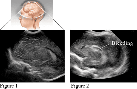
The transfocal ultrasound (or transcranial ultrasound) uses reflected sound waves to record images of the skull and the chambers containing the cerebrospinal fluid inside (the ventricles). This method is often performed in neonates to evaluate complications related to the brain, especially with premature babies.On the other hand, ultrasound cannot penetrate bones, so transfocal ultrasound cannot be performed when the baby's skull bones have formed and developed. Thus, transthoracic ultrasound is only performed for infants, children under 18 months of age, breastfed children (when the skull bones are not really complete) or in adults before brain surgery (when the skull bones are not fully developed). is opened). In particular, transfocal ultrasonography is often performed to evaluate intracranial and ventricular problems in children and infants, including ventricular dilatation.
Transthoracic ultrasound helps to assess the size of the newborn's head, evaluate the dilatation of the lateral ventricles, the dilation of the right ventricle, and the dilation of the left ventricle. In addition, this type of ultrasound can help detect inflammation in or around the skull (such as encephalitis or meningitis), and screen for congenital problems of the skull (such as hydrocephalus).
3. When to perform transfocal ultrasound?
In addition to diagnosing ventricular dilatation in infants, transfocal ultrasound is also used to:Evaluate hydrocephalus; Detecting bleeding brain tissue or ventricles; Determine whether there is damage to the white matter around the ventricles (white matter injury) or not; Detect some birth defects; Locate the site of infection or tumor.

4. What preparation is needed before performing transfocal ultrasound?
The doctor can outline the steps to be taken for parents or children before performing transfocal ultrasound.For relatively large pediatric patients, when performing an ultrasound, it can be easy to become hungry. In this case, you can feed your baby to help him feel more comfortable and stay still during the ultrasound.
5. Procedure for performing transthoracic ultrasound
To diagnose an infant with dilated ventricles, the doctor will ask the parent to keep the baby still during the ultrasound. The sonographer will then apply a gel on your baby's scalp right at the fontanelle (where the skull bones come into contact, but have not yet fully closed). The function of this gel is to help propagate sound waves.Next, the sonographer will place a small probe on the baby's head that is connected to a computer system, and then move it to many places on the gel. This transducer sends high-frequency sound waves into the ventricles and back. The computer system then records the sound waves and converts them into images. The doctor will rely on this image to analyze and diagnose the child with dilated ventricles.
6. What should be done when the child has dilated ventricles?
After the ultrasound, the child can return to normal life. The doctor will rely on the ultrasound results to consider the appropriate treatment option, or order monitoring and perform additional tests.There are many cases of mild ventricular dilatation in infants, the child's body will adjust itself so that the ventricular fluid does not increase or even decrease. Therefore, when parents notice that their children have dilated ventricles, they should not be too worried, but should listen to specific advice and advice from the doctor, have regular check-ups to monitor the condition of the ventricles. detect if there are early symptoms for surgical treatment when necessary.
As a key area of Vinmec Health system, Pediatrics Department always brings satisfaction to customers and is highly appreciated by industry experts with:
Gathering a team of top doctors and nurses in Pediatrics : consists of leading experts with high professional qualifications (professors, associate professors, doctorates, masters), experienced, worked at major hospitals such as Bach Mai, 108.. Doctors All doctors are well-trained, professional, conscientious, knowledgeable about young psychology. In addition to domestic pediatric specialists, the Department of Pediatrics also has the participation of foreign experts (Japan, Singapore, Australia, USA) who are always pioneers in applying the latest and most effective treatment regimens. . Comprehensive services: In the field of Pediatrics, Vinmec provides a series of continuous medical examination and treatment services from Newborn to Pediatric and Vaccine,... according to international standards to help parents take care of their baby's health from birth to childhood. from birth to adulthood Specialized techniques: Vinmec has successfully deployed many specialized techniques to make the treatment of difficult diseases in Pediatrics more effective: neurosurgery - skull surgery, stem cell transplantation. blood in cancer treatment. Professional care: In addition to understanding children's psychology, Vinmec also pays special attention to the children's play space, helping them to have fun and get used to the hospital's environment, cooperate in treatment, improve the efficiency of medical treatment. Master. Doctor. Nguyen Minh Tuan has over 27 years of experience in the field of Pediatrics. The doctor was former Deputy General Department of Pediatrics - Da Nang Obstetrics and Gynecology Hospital before being a Pediatrician at the Department of Pediatrics - Neonatology - Vinmec Danang International General Hospital as it is now
Please dial HOTLINE for more information or register for an appointment HERE. Download MyVinmec app to make appointments faster and to manage your bookings easily.


