This is an automatically translated article.
The article was professionally consulted by Doctor Tran Hai Dang - Deputy Head of Radiology Department - Department of Diagnostic Imaging - Vinmec Central Park International General Hospital.CT brain perfusion is an advanced technique used in many developed countries around the world. In Vietnam, this technique is also applied in many large hospitals in stroke cases. Thereby helping to accurately diagnose and provide quick and effective treatment for the patient.
1. Learn about brain perfusion CT scan technique
CT scan, also known as computed tomography (CT scan) is a technique that uses a computer and X-ray machine to create cross-sectional images of the body. From these images will provide more detailed information than conventional x-ray. From the CT scan, the doctor will clearly see the soft tissues, blood vessels and bones in many different parts of the body.

Mô phỏng chụp CT mạch máu não
CT brain perfusion method uses imaging techniques to survey perfusion of brain parenchyma. There will be a scanner to take pictures of the blood vessels, which will be connected to a computer, from which 2D and 3D images will be displayed on the screen. With the information obtained, doctors will assess the extent of brain parenchymal damage in stroke diseases, clearly identify the area of ischemic brain tissue that can still be saved. From there, doctors make quick and appropriate treatment decisions. In addition, cerebral perfusion CT can also evaluate vascular abnormalities in dilated, narrowed, and occluded intracranial and extracranial vessels.
During a CT scan of the brain, doctors will use contrast dye injected into a vein according to a special examination procedure to clearly see the cerebral blood vessels, measure the cerebral blood flow and evaluate the lesions. brain parenchymal injury.
2. What should be noted before CT brain perfusion scan?
CT perfusion can have some risks such as natural radiation contamination (if present, it is also below the level that causes adverse effects on the body) as well as possible allergy to contrast dye... but here is a very important examination to help diagnose diseases that are often related to the patient's life, so it is often prescribed by doctors in emergency and in specialist visits.
Therefore, before taking a CT angiogram, doctors need to check the following information of the patient:
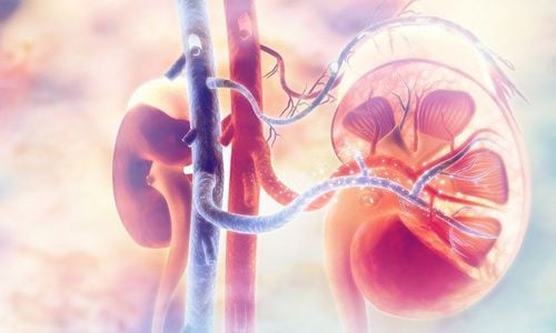
Pregnant or breastfeeding Have had an allergic reaction to contrast in a previous scan History of kidney disease Asthma and daily medication Diabetic being treated with Metformin History of hyperthyroidism

Chụp CT ở phụ nữ đang mang thai và cho con bú cần được các bác sỹ chuyên khoa cân nhắc kỹ lưỡng giữa lợi ích-nguy cơ và được bảo vệ an toàn bức xạ nếu như bắt buộc phải chụp.
3. CT angiography is useful in diagnosing stroke
CT cerebral perfusion is a non-invasive technique, very useful in surveying and diagnosing emergency diseases related to cerebral blood vessels. If a patient is brought to the emergency hospital with suspicion of cerebral infarction, this technique will be applied first to determine the cause and extent of cerebral infarction, to distinguish between infarction and hemorrhage.. From there, the most suitable course of treatment can be established. CT brain perfusion is the preferred use in stroke cases because it provides a quick and accurate diagnosis and helps to make timely treatment decisions.
This technique can also be used and is very useful in screening people with suspected cerebrovascular diseases such as cerebral aneurysms, cerebrovascular stenosis/occlusion.
With proactive screening for cerebrovascular diseases, each person will reduce the risk of stroke, sudden death, and the sequelae left by stroke. Thereby reducing the burden on family and society.

Chụp CT tưới máu não
4. Brain perfusion CT scan at Vinmec Central Park International General Hospital for early diagnosis of stroke.
To serve the most effective examination and treatment for those at risk or who have had a stroke, the Diagnostic Imaging Department - Vinmec Central Park International Hospital has been using CT scanning technique. Cerebral perfusion is used to diagnose ischemic and hemorrhagic lesions, to help guide timely and effective treatment, as well as to help screen the risk of stroke in some cerebrovascular diseases.
CT brain perfusion technique at Vinmec Central Park International General Hospital is performed with a success rate of over 95%.

Hệ thống máy MSCT 640 lát cắt hiện đại tại Vinmec
With a modern system of 640-slice MSCT machines and the world's leading synchronous and advanced equipment, with a team of highly qualified and experienced doctors - technicians, imaging department Imaging can perform many diverse and specialized techniques, supporting clinicians to diagnose diseases quickly and accurately, and help monitor disease during and after treatment.
BS. Tran Hai Dang - Diagnostic Imaging Department - Vinmec Central Park International General Hospital. The doctor has long trained abroad with certificates of continuous training, postgraduate training in diagnostic imaging and intervention in Switzerland, the United States, Austria, France, and participated in many international conferences. The international conference on advanced diagnostic imaging is held in countries with advanced medicine in the world. With the experience and success that he has built up over the past 30 years, Dr. Tran Hai Dang has been trusted by many patients to register for treatment at Vinmec.
Please dial HOTLINE for more information or register for an appointment HERE. Download MyVinmec app to make appointments faster and to manage your bookings easily.