This is an automatically translated article.
The article is professionally consulted by Master, Doctor Nguyen Thuc Vy - Doctor of Diagnostic Imaging - Department of Diagnostic Imaging - Vinmec Nha Trang International General Hospital.There are many diseases of the spleen that can be identified through ultrasound. Spleen ultrasound is simple, quick and painless.
1. What is a spleen?
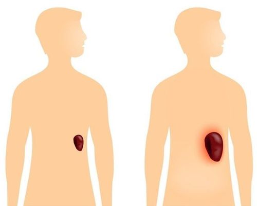
The spleen is an internal part of the abdominal cavity of the body, when observing this part through imaging means, the position of the spleen will be in the left corner. The size of the spleen is only approximately 150-200g for a normal adult. The structure of the spleen consists of a shell and a part of the spleen parenchyma.
The spleen has the function of filtering blood for the body, through the blood filtration process to proceed to remove all red blood cells that are defective, old or have poor function. In addition, the spleen also has a white marrow system, which helps create antibodies to protect the body.
2. Diseases of the spleen
The following are some diseases of the spleen that can be identified through ultrasound.
2.1 Strong spleen
Hypersplenism is a phenomenon in which the spleen does not filter, but destroys all the red blood cells that pass through or often suddenly destroys a large amount of blood in the body.
Hyperthyroidism often occurs in patients with blood cancer, liver disease or an infection of the body caused by a certain bacteria.
Some cases lead to hyperthyroidism because this part has to work in an overloaded way, causing inherent dysfunction.

Bệnh nhân ung thư máu có thể bị cường lách
2.2 Contusion or rupture of the spleen
This pathology can be easily detected through ultrasound of the spleen. The cause of spleen rupture is due to a strong accident, because malaria bacteria swell the spleen causing the outer protective covering of the spleen to burst.
2.3 Spleen infection
Infection of the spleen can also be easily checked through ultrasound. Currently, the most commonly reported spleen infections include two types of infections: bacterial splenic abscess and fungal splenic abscess.
2.4 Cancer of the spleen
Common spleen cancer will be of two types, primary spleen cancer and secondary spleen cancer. Cases of cancer of the spleen are quite rare and an ultrasound scan can detect growths in this area. Some cases of spleen cancer were recorded without tumors, but only the spleen was larger than normal when conducting ultrasound.

Ung thư lá lách rất hiếm gặp
3. Why is an ultrasound of the spleen needed?
Although the spleen is a relatively small organ compared to the size of the body, it plays an important role and is located in the human immune system. However, because it is quite small in size and thin and strong, it is easy to be crushed if it encounters a strong impact with the body or breaks naturally because of the appearance of devastating diseases.
Therefore, in addition to the cases with obvious and indicated signs, it is also necessary to conduct a periodic ultrasound of the spleen to check the development of abnormalities of this organ
4. Indicated cases of spleen ultrasound
In order to detect the above-mentioned diseases of the spleen, the doctor will appoint ultrasound for the following cases:
Verify splenomegaly and monitor the progress of the spleen through treatment. Detecting splenomegaly with uniform, diffuse or regional echogenicity, thereby inferring the pathology of the spleen. Detection of hematoma or splenic rupture after trauma.

Hình ảnh vỡ lách
5. Steps to perform ultrasound diagnosis of spleen pathology
5.1 Patient preparation
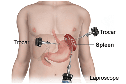
With the form of spleen ultrasound, the patient will be asked to fast for clearer images. At the same time, baryte should not be used for gastrointestinal imaging and laparoscopy should not be used.
5.2 Techniques used
Spleen ultrasound will use a 3.5MHz or 2.25MHz transducer (in obese patients). During the ultrasound, the patient will breathe out and hold his breath, when necessary, the patient can drink water to separate the spleen from the liver (T) lobe.
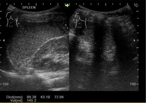
Hình ảnh lá lách trên siêu âm
5.3 Steps to take
The spleen ultrasound process will be relatively quick and painless. When entering the ultrasound process, the patient will be asked by the doctor to lie on his or her back or to the right depending on the location of the pain or the condition of the disease. In some special cases, it may be necessary to switch to a sitting position.
During the ultrasound, the doctor may order the patient to do some other tests to check the position of the abdominal cavity. This general examination step will help the doctor more accurately assess the specific health of each patient and come up with a more appropriate and effective treatment plan.
Conducting an ultrasound of the spleen can accurately identify diseases of this part. Despite its small size, it plays an extremely important role in human health. Therefore, periodic ultrasound of the spleen is recommended by doctors to determine the status of the spleen and detect early diseases if any.
Master. Doctor Nguyen Thuc Vy has 09 years of experience in Diagnostic Imaging. Dr. Vy has many years of experience working in the Department of Diagnostic Imaging at the University of Medicine and Pharmacy Hospital in Ho Chi Minh City, trained and attended many courses on specialized imaging at the University of Medicine and Pharmacy Hospital. Hue Pharmacy, Ho Chi Minh City University of Medicine and Pharmacy, Cho Ray Hospital. Currently working at the Diagnostic Imaging Department of Vinmec Nha Trang International General Hospital
Please dial HOTLINE for more information or register for an appointment HERE. Download MyVinmec app to make appointments faster and to manage your bookings easily.