This is an automatically translated article.
A thorough understanding of the risks and progression of diabetic retinopathy will help you choose the right treatment for yourself before they cause more serious complications.
1. What is diabetic retinopathy?
Diabetic retinopathy is an eye condition that can lead to abnormal changes to the blood vessels in the retina.
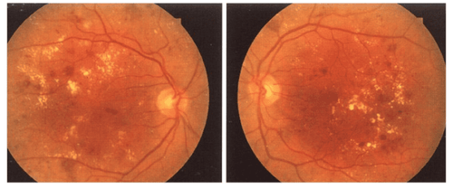
The retina is a lining at the back of the eye that converts light into images. With diabetic retinopathy, the blood vessels in the eye swell, causing fluid to leak or bleed, leading to dangerous changes in vision and even blindness. Diabetic retinopathy often affects both eyes, severely impairing vision. If not treated early, this disease can leave scars and damage the patient's retina.
Currently, diabetic retinopathy is considered the leading cause of vision loss for people with diabetes. Therefore, you need to learn carefully the causes, signs, as well as how it develops into a serious eye problem, in order to prevent and treat the disease effectively.
2. Common Symptoms of Diabetic Retinopathy
In its early stages, diabetic retinopathy usually doesn't have any noticeable symptoms. However, when it is severe, the disease can cause some common symptoms such as:
Blurred vision ; Inability to see colors, impaired vision; Loss of central vision; There are holes or black spots in your vision; Floating spots appear in your vision.
3. What causes diabetic retinopathy?
The cause of diabetic retinopathy begins when your blood sugar level is too high for a long time, blocking the small blood vessels that protect the retina of the eye. At this point, the eye will have to try to grow new blood vessels, but these blood vessels grow abnormally and begin to weaken, which can leak blood and fluids into your retina, causing eye complications of diabetes.
In addition, diabetic retinopathy can lead to another condition known as macular edema. When macular edema becomes severe, more blood vessels become blocked. Scar tissue begins to form as new blood vessels in the eye have grown. This extra pressure can cause the retina to tear or detach, increasing the risk of blindness in people with diabetic retinopathy.
Any form of diabetes, including type 1 , type 2 and gestational diabetes can develop into diabetic eye complications. The risk increases as diabetes persists for many years.
Several other factors can also increase the incidence of diabetic retinopathy including:
High cholesterol levels in the blood; High Blood Pressure; Smoking cigarettes regularly; Be Hispanic, African-American, or Native American.

Võng mạc tiểu đường được xem là nguyên nhân hàng đầu gây mất thị lực cho những người mắc bệnh tiểu đường
4. How many stages of diabetic retinopathy?
Diabetic retinopathy is divided into 4 main stages, including:
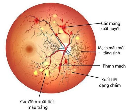
4.1 Stage 1: Mild non-proliferative retinopathy This is the first stage of diabetic retinopathy, also known as retinopathy. background. During this stage, the blood vessels in the retina have developed some small bulges. These bulges, called aneurysms, can cause blood vessels to leak a small amount of blood into the retina.
In the first stage, the patient may not have any vision problems, so no treatment is required. However, each person should still talk to their doctor about measures to help keep diabetic retinopathy from getting worse. At the same time, actively control your blood pressure, blood sugar and cholesterol levels.
Planning for screening tests in 12 months is a must. If the condition progresses in both eyes, there is an approximately 25% chance of progressing to the third stage of the disease within the next 3 years.
4.2 Stage 2: Moderate Non-Proliferative Retinopathy The second stage is also known as pre-proliferative retinopathy. During this stage, the blood vessels in the retina swell and cannot carry blood as efficiently as before. This can cause physical changes to the retina and cause diabetic macular edema (DME).
According to research, half of people with diabetic retinopathy will develop macular edema, it can occur at any stage, but the risk is higher as the diabetic retinopathy progresses.
In the pre-proliferative stage of the retina, it also means that the disease will affect vision more. Your doctor may recommend that you have your eyes checked every 3 to 6 months.
4.3 Stage 3: Severe non-proliferative retinopathy This stage is also known as proliferative retinopathy. At that time, the blood vessels will become more congested than before, causing less blood to reach the retina and forming scar tissue. The lack of blood triggers signals to the retina to create new blood vessels. If these blood vessels are completely closed, it results in a condition known as macular ischemia. This can cause a person's vision to become blurred and dark spots, also known as flies, appear in front of the eyes.
In stage 3, the patient will have a very high risk of partial vision loss. Prompt treatment at this time will help prevent total vision loss. However, it is also important to note that a part of vision that is lost cannot be restored.
4.4 Stage 4: Proliferative diabetic retinopathy (PDR) This is the most advanced stage of the disease, new blood vessels grow in the retina and form gel-like fluids that fill the eye. This growth is called neovascularization. These new vessels are usually very thin and weak, and can bleed and form scar tissue. When scar tissue begins to form, it can pull the retina away from the tissue underneath the patient's eye, leading to retinal detachment. This condition can cause permanent loss of both straight and side vision (permanent blindness).
5. Diagnosing Diabetic Retinopathy
To diagnose diabetic retinopathy, your doctor will conduct an eye exam to check your ability to see at different distances. At the same time, the pressure inside the eye can be checked and the patient uses eye drops that dilate the pupils to look for abnormal changes in the blood vessels and to see if any new blood vessels are growing. Check to see if the retina is swollen or detached?
In addition to the eye exam, the doctor will have the patient perform an optical tomography (OCT) scan. This method uses light waves to take pictures of the inside of the eye and look for signs of disease.
The final test to diagnose diabetic retinopathy is fluorescein angiography. This technique will help detect diabetic macular edema (DME) or eye complications of severe diabetes. The doctor will inject dye into a vein (usually in the arm), then the dye will travel to the eye and the doctor can see an image of the blood vessels in the retina, thereby helping to detect Leaks and eye damage.

Giai đoạn đầu của bệnh võng mạc tiểu đường còn được gọi là bệnh võng mạc nền
6. Treating Diabetic Retinopathy
The treatment of diabetic retinopathy is usually based on the level and health status of the patient, some methods may be indicated including:
Anti-VEGF therapy: This is drug therapy to prevent Blocks vascular endothelial growth factor (VEGF), which helps reverse the growth of blood vessels and reduces fluid accumulation in your retina. Anti-VEGF drugs commonly used to treat diabetic retinopathy include: bevacizumab (Avastin), aflibercept (Eylea), and ranibizumab (Lucentis). Laser surgery: Lasers can create small burns over areas of leaky blood vessels in the macula of the eye. People with diabetic retinopathy may need anti-VEGF therapy after this surgery. Dispersive laser surgery: This treatment can create up to 2,000 small burns to treat spots where the retina has separated from the macula. Dispersive laser surgery can help shrink abnormal blood vessels in the retina and save central vision in people with diabetic retinopathy. This treatment is usually most effective if the patient has it before the new vessels start to bleed. Using corticosteroids: Specialist doctors can implant or inject short-term and long-term corticosteroids into the eyes of diabetic retinopathy patients for treatment. However, these drugs can increase the risk of cataracts and glaucoma, so it is important to monitor your eye pressure regularly while using them. Removal of vitreous in the eye: You will need to have the vitreous (crystal) removed from the eye if blood vessels leak into the retina and vitreous. This procedure will remove blood leaking into the retina, help patients see better, and effectively treat blurred vision.
7. Prevent Diabetic Retinopathy
To prevent and slow the progression of diabetic retinopathy, patients need to
See an ophthalmologist at least once a year for a comprehensive eye exam; If you have diabetes and are pregnant, you should have regular eye exams during the first 3 months of pregnancy; Do not smoke if you have diabetes or diabetic retinopathy. In case you want to know how much blood sugar is, what level of diabetes is, patients can sign up for a diabetes screening package, dyslipidemia at Vinmec International General Hospital. Not only health screening, the diabetes and dyslipidemia screening package also helps to detect pre-diabetes early, accurately classify diabetes type, develop a nutritional regimen, and monitor the treatment of diabetes. risks and complications caused by diabetes.
Using the screening package for diabetes and dyslipidemia at Vinmec, customers will receive:
Endocrine examination (with appointment); Total urinalysis (by automatic machine); Glucose quantification; Quantification of HbA1c; Quantification of Uric Acid; Quantification of Cholesterol; Quantification of HDL-C (High density lipoprotein Cholesterol); Quantification of LDL-C (Low density lipoprotein Cholesterol); Triglyceride quantification; Quantification of Urea; Quantification of Creatinine; Measure AST activity (GOT); Measure ALT activity (GPT); Measure GGT activity (Gama glutamyl Transferase); Quantification of MAU (Micro Albumin Arine); Echocardiography, transthoracic pericardium; Normal electrocardiogram; Doppler ultrasound of the carotid artery, transcranial (carotid) Doppler; Doppler ultrasound of the arteries and veins of the lower extremities (bilateral lower extremity arteries).
Please dial HOTLINE for more information or register for an appointment HERE. Download MyVinmec app to make appointments faster and to manage your bookings easily.
Reference sources: modernod.com, webmd.com


