This is an automatically translated article.
Epithelial polyps are the most common type of gastric polyp. These include basal glandular polyps (FGPs), hyperplastic polyps, and adenomatous polyps. Other less common epithelial lesions that may present as polyps include neuroendocrine tumors (formerly known as carcinoids), ectopic pancreatic tissue, and pyloric adenomas.
1. Gastric neuroendocrine tumors (formerly known as carcinoids)
Most gastric neuroendocrine tumors occur in the body and fundus of the stomach (90%), stained with chromogranin A or synaptophysin by immunohistochemistry.
There are four types of gastric carcinoids, each arising in different clinical settings, and each with its own distinct prognosis and treatment regimen. This particular tumor emphasizes the importance of parallel biopsies of the basal adipose tissue.
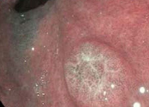
On endoscopic images, they appear as submucosal mass lesions, sometimes with ulcers.
In type I and II carcinoids, some polyps are seen in clusters, arising almost exclusively in the body's mucosa. Type III lesions are usually solitary and may present throughout the stomach. Class IV carcinoids can arise anywhere in the stomach and have a significantly worse prognosis. Histologically, these neuroendocrine tumors appear to be similar, and biopsy of the nonpolypoid mucosa is important in differentiating tumor type, prognosis, and modality.
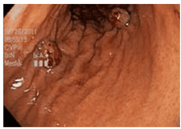
U thần kinh nội tiết dạ dày (trước đây gọi là carcinoid)
2. Ectopic pancreatic tissue
Ectopic pancreatic tissue is pancreatic tissue that lacks anatomical and vascular continuity with the main organ of the pancreas. Usually located in the stomach, these lesions are very small and are discovered incidentally.
Endoscopically, they appear as submucosal masses and may be misinterpreted as other submucosal tumors (ie, gastrointestinal stromal tumors [GISTs] or lipomas leiomyomas) . The umbilicus at the center of this structure is often considered the endoscopic clue to confirm the diagnosis of these lesions, although histologically demonstrable ectopic pancreatic tissue may be seen without central concave sign on this endoscope. These lesions have a particularly low malignancy potential.
3.Ultra pyloric
Pyloric adenoma is a rare tumor demonstrating differentiation of the gastric epithelium. They are composed of pyloric, closely spaced tubules with a single layer of low columnar to monolayer epithelial cells.
They usually arise in the stomach with basal mucosa, show features of autoimmune gastritis and intestinal metaplasia, and present with a female predominance. They can often be confused with abnormal gastric hyperplastic polyps; they are distinguished by displaying a thin ground glass cytoplasm devoid of apical mucin.

Hình ảnh vị trí môn vị
4.Hamartomatous Polyp
Cellular polyps are usually mucosa-based but can originate in any of the three embryonic layers. These polyps are the result of disordered growth of local tissues.
Examples include Peutz-Jeghers polyps and juvenile polyps, as well as hamartomatous polyps with no specific name. They may be of a sporadic nature or be associated with various multiple polyposis syndromes such as Peutz-Jeghers syndrome, juvenile polyposis syndrome, and Cowden syndrome (PTEN or multiple hamartoma syndrome), as discussed. under.
5.Polyp Peutz-Jeghers
Peutz-Jeghers syndrome is an autosomal dominant disorder with a unique set of findings, including lipomas throughout the gastrointestinal tract and hyperpigmentation, especially in the gastrointestinal tract. each.
Peutz-Jeghers polyps have a characteristic glandular epithelium with dilated follicular glands, supported by a skeleton of well-developed smooth muscle adjacent to the muscular mucosa. In the small intestine and colon, these lesions can be distinguished from juvenile polyps, as Peutz-Jeghers polyps have an intact stroma with a "fan-shaped" appearance of smooth muscle fibers.
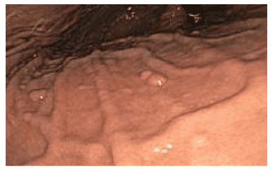
Polyp Peutz-Jeghers là một tập hợp con của các polyp dạng hamartomatous.
Peutz-Jeghers polyps are potentially malignant and the mean age of patients with gastric carcinoma is estimated to be 30 years.
Current recommendations recommend screening for gastric polyps as early as 8 to 21 years of age. Peutz-Jeghers polyps in the stomach larger than 1 cm should be resected endoscopically and the patient should be monitored annually. For patients with smaller (<1cm) polyps, endoscopic surveillance is recommended every 2 to 3 years, although it is recognized that small polyps can be removed in certain medical facilities.
6. Juvenile Polyposis and Juvenile Polyposis Syndrome
Juvenile polyps are mucosal neoplasms consisting mainly of stroma and dilated cystic glands; they are therefore classified as hamartomatous polyps.
They are sometimes called inflammatory polyps or polyps due to the appearance of swollen, mucus-filled glands, inflammatory cells, and edema. Juvenile polyps are usually solitary lesions of the scalp and range in size from 3mm to 20mm. When found singly, they are thought to be benign, non-syndromic random lesions.
Juvenile polyps are an autosomal dominant disorder that carries a 50% higher lifetime risk of gastric malignancy. Therefore, for juvenile polyposis syndrome, endoscopic screening is recommended starting at age 18 and every 3 years thereafter.
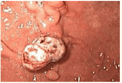
Polyp vị thành niên
7. Cowden's syndrome
Cowden's syndrome is a multisystem, autosomal dominant disorder characterized by overgrowth of hamartomatous tissue of all three embryonic layers.
80% of patients have germline mutations of the PTEN tumor suppressor gene.
There are several prognostic criteria for the diagnosis of this syndrome, including facial trichilemmoma, keratosis, and papilloma.
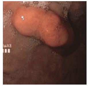
Hội chứng Cowden
Cowden syndrome polyps are a subset of many stromal polyps. Gastrointestinal polyps are generally benign in nature. There are associated abnormalities of the breast (carcinoma), thyroid (follicular) and genitourinary (endometrial carcinoma) systems. These hamartomas are the least specific and may contain varying degrees of mixed tissue types, such as mixed glands, vessels, and smooth muscle.
Please dial HOTLINE for more information or register for an appointment HERE. Download MyVinmec app to make appointments faster and to manage your bookings easily.


