This is an automatically translated article.
Gastric polyps usually originate in the gastric mucosa; In about 6% of cases found during upper GI endoscopy, gastric polyps are a heterogeneous group of epithelial and subepithelial lesions. Most have no risk of cancer, but there are a small number of polyps that are potentially malignant, requiring further endoscopic treatment or periodic monitoring.1. Mesenchymal is what part of the stomach?
Mesenchymal is the term for the developing sparse connective tissue of the embryo, which mainly originates in the mesoderm and gives rise to the majority of connective tissue cells in the adult body.
2. Polyps originate from the stomach's mesenchyme
Over 90% of polyps arising from the gastric mesenchyme are usually uncomplicated, only larger polyps may present with bleeding, anemia, obstruction, or abdominal pain.

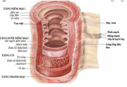
Hình 1: Cấu tạo thành ống tiêu hoá
2.1 Epithelial polyps
Epithelial polyps are the most common type of gastric polyp. These include basal glandular polyps (FGPs), hyperplastic polyps, and adenomatous polyps. Other less common epithelial lesions that may present as polyps include neuroendocrine tumors (formerly known as carcinoids), ectopic pancreatic tissue, and pyloric adenomas.
2.2 Mesenchymal polyps
Mesenchymal lesions include a broad spectrum of tumors of dermis origin. These polyps can be mucosal or submucosal but are usually located below the surface epithelium, forming a nodule rather than a polyp.
Given their deep location, these lesions should be further evaluated by endoscopic ultrasound (EUS) and tissue collection.
2.3 Inflammatory fibroid polyps (abbreviated as IFP - Inflammatory fibroid polyps)
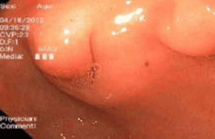
Inflammatory polyps usually present as polypoid lesions or sessile nodules and are small (<1.5 cm). They are well-described lesions located in the posterior thoracic or anterior thoracic region. Microscopic examination shows that this lesion consists of vessels, small rhabdoid cells (circled) and collagen fibers (arrows), as well as an inflammatory background with numerous eosinophils.

Hình 2: Polyp dạng sợi viêm.
These polyps also contain myxoid stroma and are usually thin-walled vessels. IFP can occur at any age but is most common between the ages of 50-60 and has a slightly higher incidence in women. These are rare lesions, with an estimated relative prevalence of 0.09%.
IFP usually presents as a solitary lesion in the pylorus of the stomach or the distal antrum and is usually small (<1.5 cm) and sessile. These lesions are considered unlikely to be malignant; therefore, endoscopic follow-up after initial histological confirmation is not recommended. Larger and/or symptomatic lesions may require complete resection by an experienced endoscope.
2.4 Gastrointestinal stromal tumor (GIST)
GISTs are rare connective tissue tumors that originate in the muscularis stroma and account for 1% to 3% of all gastrointestinal malignancies. GISTs are often found incidentally during upper endoscopy performed for reasons or symptoms unrelated to the tumor.
Clinical features may vary depending on the anatomical location, size and aggressiveness of the tumor. Although these tumors can arise anywhere along the digestive tract, the most common site is the stomach.
Tumor treatment depends on the stage of the tumor. If localized to the stomach, the tumor can be surgically removed. If the tumor is metastatic or cannot be removed.

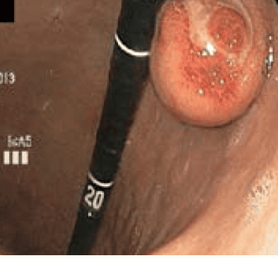
Hình 3: Các khối u mô đệm đường tiêu hóa (GIST).
GISTs are subepithelial polyps that can arise from any site in the gastrointestinal (GI) tract. They are often accompanied by an ulcerated center. These lesions arise from the interstitial cells of the Cajal, the pacemaker cells of the gastrointestinal tract located between the inner and outer longitudinal layers of the muscularis stroma (MP).
2.5 Leiomyomas
Leiomyomas are benign smooth muscle tumors. Leiomyomas are often asymptomatic and discovered incidentally. Endoscopically, they appear as circular submucosal lesions with intact upper mucosa. To the touch, they feel like rubber.
2.6 Granulomatous cell tumor
Granulomatous cell tumors are usually found in the esophagus and occur between the ages of 40 and 60 years. Endoscopically, they appear as yellow subepithelial masses or nodules, often mistaken for lipomas, although they are firmer on biopsy. Histologically, the tumors arise in the submucosal layer. Most are benign, but malignancy has been reported.
3. Conclusion
Gastric polyps are a common finding during routine endoscopy. 90% asymptomatic and unlikely to be malignancy, a subset of gastric polyps requires further intervention and histological evaluation to determine polyp type and presence of dysplasia.
Vinmec International General Hospital is one of the hospitals that not only ensures professional quality with a team of doctors, modern equipment and technology, but also stands out for its examination, consulting and service services. comprehensive and professional medical treatment; civilized, polite, safe and sterile medical examination and treatment space. Customers who choose to perform tests and treat diseases here can be completely assured of the accuracy and high efficiency in the treatment process.
Please dial HOTLINE for more information or register for an appointment HERE. Download MyVinmec app to make appointments faster and to manage your bookings easily.


