This is an automatically translated article.
This article was professionally consulted by a Doctor of Radiology, Department of Radiology - Vinmec Central Park International General Hospital.Transesophageal echocardiography not only captures specific images but also provides a lot of detail. From the small details, the doctor can conclude about the problems that need treatment.
1. What is Esophageal Echocardiography?
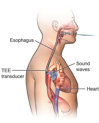
An echocardiogram is a diagnostic test that uses high-frequency ultrasound waves to obtain dynamic images of the heart and its related structures. Depending on the patient's condition and pathological progress, the doctor will prescribe the most appropriate echocardiographic method.Transesophageal echocardiography is a method that uses ultrasound waves and a transducer to be inserted through the mouth into the esophagus to detect cardiovascular diseases, because the esophagus and heart chambers are located next to each other. When it comes into contact with the esophagus, it has reached a special position close to the heart.
Ultrasound waves will be emitted to the heart chambers and then reflected back so that the doctor can get an image of the inner parts of the heart. With this method, ultrasound waves will not affect the patient's health, but also obtain the most detailed images compared to external ultrasound methods such as transthoracic ultrasound.
Siêu âm tim qua thực quản là phương pháp sử dụng sóng siêu âm và đầu dò để đưa qua đường miệng vào trong thực quản nhằm phát hiện các bệnh lý về tim mạch
2. When should transesophageal echocardiography be performed?
Transesophageal echocardiography not only captures specific images but also provides a lot of detail. From the small details, the doctor can conclude about the problems that need treatment. Transesophageal echocardiography should be performed when:The patient cannot apply conventional echocardiographic methods or the ultrasound results are not specific. People with thick chest wall due to physical location or overweight or obese. Transesophageal echocardiography when the patient is having to use wound dressings and specialist bandages to treat pathologies. Indications for transesophageal echocardiography when the patient has just performed surgery directly related to the heart. When a patient has a pericardial infection or pericardial effusion, transesophageal echocardiography must be performed, then the indications for convalescent treatment should be performed.

Phương pháp siêu âm tim qua thực quản nên thực hiện khi người bệnh có thành ngực dày do cơ địa hoặc thừa cân, béo phì
3. Some commonly used slices in esophageal echocardiography
When doing esophageal ultrasound, some commonly used slices by doctors include:Long axis right ventricular receiving chamber view: What is the esophageal echocardiogram used for 2D ultrasound, color Doppler spectrum of the tricuspid valve, Continuous transvalvular Doppler to measure peak velocity through the tricuspid regurgitation spectrum. Short axis view: This section is commonly used when 2D transmitral level ultrasound, aortic and tricuspid valve coloration, color Doppler of aortic valve, transverse ventricular transverse myocardial and transverse valvular aorta, continuous Doppler across the tricuspid valve. Short axial cross-section for maximum velocity measurement across the tricuspid regurgitation spectrum. 2-chamber view from the apex: Used when mitral valve color Doppler spectrum, 2D ultrasound. 3-chamber view from the apex: Used when 2D ultrasound, color Doppler spectrum of mitral valve, continuous Doppler spectrum across the aortic valve, pulse Doppler spectrum across the aortic valve and color spectrum of the aortic valve. 3-chamber view from the apex used to measure differential pressure across the aortic valve, left ventricular outflow tract VTI. 4-chamber view from the apex: On 2D ultrasound, the Doppler spectrum is continuous through the mitral and tricuspid valves, and the color Doppler spectrum through the mitral and tricuspid valves. 4-chamber view from the apex to measure mean differential pressure across the mitral valve, maximal flow velocity of tricuspid regurgitation. 5-chamber view from the apex: This esophageal echocardiogram is used when 2D ultrasound, pulse Doppler spectrum through the left ventricular outflow tract, color Doppler spectrum of the aortic valve and continuous Doppler spectrum across the aortic valve. 5-chamber view from the apex to measure differential pressure across the aortic valve, left ventricular outflow VTI. Parasternal long axis: This slice is often used when 2D ultrasound, VHL color Doppler ultrasound, aortic valve. Simultaneously to measure parameters such as left ventricular outflow tract, left ventricular end-diastolic diameter, interventricular septal thickness or left ventricular posterior wall thickness and used to measure Valsalva sinus diameter. Subcostal view: When using 2D ultrasound, measuring inferior vena cava diameter, respiratory collapse of inferior vena cava, using subcostal view. Upper sternal view: Used when 2D ultrasound of the aortic arch.
4. Attention when transesophageal echocardiography

Trước khi siêu âm thực quản, người bệnh cần nhịn ăn 6 tiếng
Before the esophageal ultrasound, the patient should: Fast for 6 hours before the ultrasound; take the medicine with a very small sip of water; Remove the denture, then lie on the left side. After being numbed, open your mouth wide and bite into the plastic jaw to protect the teeth and the tube. After the ultrasound: The patient can eat and drink at the earliest 1 hour after the ultrasound. Because sedation is still effective, alcohol should not be consumed 1-2 days after the procedure. The anesthetic will last for this time. In summary, transesophageal ultrasound technology contributes to improving the treatment efficiency of complex cardiovascular diseases. Moreover, transesophageal echocardiography does not cause pain and discomfort to the patient, so you can rest assured to perform this echocardiographic therapy at reputable hospitals across the country. . Currently, Cardiovascular Center - Vinmec Times City International General Hospital is the address for quality basic echocardiography. This is one of the leading centers in the country for examination, diagnosis, screening and treatment of cardiovascular diseases. With the convergence of a team of experienced and reputable experts in the field of surgery, internal medicine, interventional cardiac catheterization and the application of advanced techniques in the diagnosis and treatment of diseases. Cardiovascular management, together with a system of modern equipment, on par with the most prestigious hospitals in the world such as: MRI 3 Tesl a (Siemens), CT 640 (Toshiba), other equipment advanced endoscopy equipment EVIS EXERA III (Olympus Japan), advanced anesthesia system Avace, Hybrid operating room according to international standards... Cardiovascular center at Vinmec Times City International General Hospital has reaped the benefits. achieved many successes and gained the trust of many patients.
Please dial HOTLINE for more information or register for an appointment HERE. Download MyVinmec app to make appointments faster and to manage your bookings easily.