This is an automatically translated article.
The article is professionally consulted by Master, Resident Doctor Nguyen Van Anh - Radiologist - Department of Diagnostic Imaging and Nuclear Medicine - Vinmec Times City International General Hospital.Today, with the great development of science and technology, people have introduced many different imaging techniques to coordinate and support in the treatment of diseases. In addition to conventional diagnostic methods such as ultrasound and X-ray, Computed Tomography Simulation (CT Sim) is also considered a "master" in monitoring and treating cancer diseases. letters.
1. What is simulated tomography?
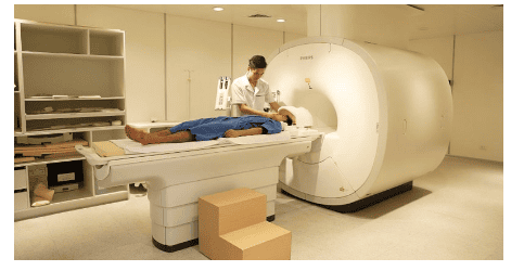
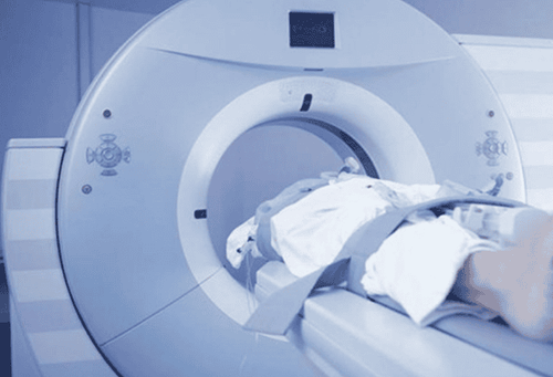
Simulated tomography (or simulated CT scan) is the same as that of conventional diagnostic tomography, this technique uses many X-rays to scan an area of the body in a cross-section, in combination with processed by computer to produce 3D images of the part to be photographed. This is a perfect combination of CT imaging and precise positioning of the simulation system.When using this technique, the patient will be simulated in a position identical to the radiotherapy treatment position, lying on a flat table surface, using appropriate immobilizers and laser positioning systems.
After the simulated tomography scan, the doctor will receive 3D image data of the patient's body part being treated. The doctor clearly sees the condition of soft tissues, blood vessels and bones in different parts of the body through these images. This is a powerful support tool for accurate planning, dosing and treatment.
Time to simulate tomography takes place within 30 minutes, longer than diagnostic CT scan. After about 1-2 weeks, the patient will begin to enter the treatment phase.

2. Application of simulation tomography technique
Thanks to all the features and features along with the precise simulation function, the simulation tomography technique is very important in cancer radiation therapy. Therefore, this is an indispensable tool, effectively supporting doctors in the work of diagnosing and treating cancer. In addition, this technique is most commonly used in radiotherapy planning software today because of its simplicity, utility and economy. From there, doctors can plan radiation therapy more accurately and simply with this simulation CT scanning technique.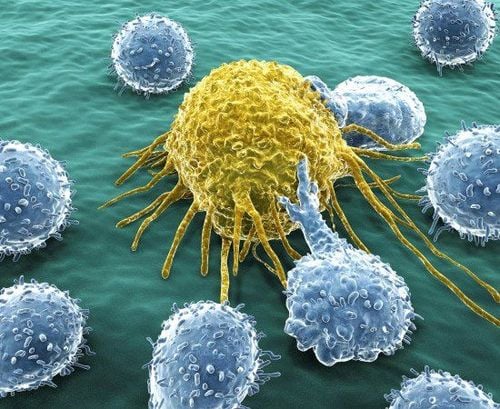
3. What you need to know about computed tomography simulation
3.1. Description of the simulated tomography technique
The tomography room will be fitted with a laser simulation system that helps to accurately locate the position, posture and coordinates when taking pictures. After capturing, simulation results will be sent to laser beam control software. This software helps us automatically calculate the distance between the tumor and the tumor center on the patient's CT scan.VPS (Virtual Planning Systems) simulation system receives CT images of patients after taking them. Through the simulation of the system, the doctor will determine the location and size of the tumor in the patient's body, which in conventional CT scanners does not have this function. The landmarks of the tumor site will be transferred to the radiotherapy accelerator system.
Accordingly, when placing the patient in the accelerator room, the laser system of the accelerator will be controlled to move to the center of the tumor based on the calculated distance above automatically, the doctor will mark the this point, precisely positioned with the marked points. The reference center of the accelerator is positioned on a line passing through the center of the tumor, and radiation therapy is performed outward from the center of the tumor. The beam will be directed at the tumor center, so the ability to kill the tumor will be enhanced because the most therapeutic dose is delivered to the tumor and minimize the dose affecting other organs.
3.2. Simulated tomography procedure
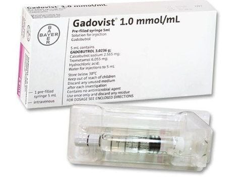
Before taking a CT simulation, the patient needs to pay attention to the following:The patient needs to fast for at least 4-6 hours before contrast injection. 2 hours before thin tissue scan can still drink a moderate amount of water. If the patient is pregnant or has one of the diseases such as diabetes, venous disease, asthma, kidney and drug allergies, it is necessary to inform the doctor immediately. All metal objects on the patient's body should be removed because they will interfere with the image when taking pictures. Accordingly, the simulation tomography procedure is conducted according to the following steps:
Step 1: The staff of the simulation CT room will check: Patient information, simulated posture, appropriate type of immobilizer . Then explain to the patient about the simulated CT scan procedure. Step 2: The patient's photo will be imported into the patient management software and pasted into the treatment sheet. Step 3: If the patient is indicated for contrast injection, an intravenous line is required. Step 4: The patient will be placed on the CT table by 2 medical staff, using an appropriate fixed pillow, adjusting the patient's position to straighten the axis in the direction of the head - feet based on the positioning laser system. To avoid the effect of the laser on their own vision, patients should keep their eyes closed during the simulation CT scan.
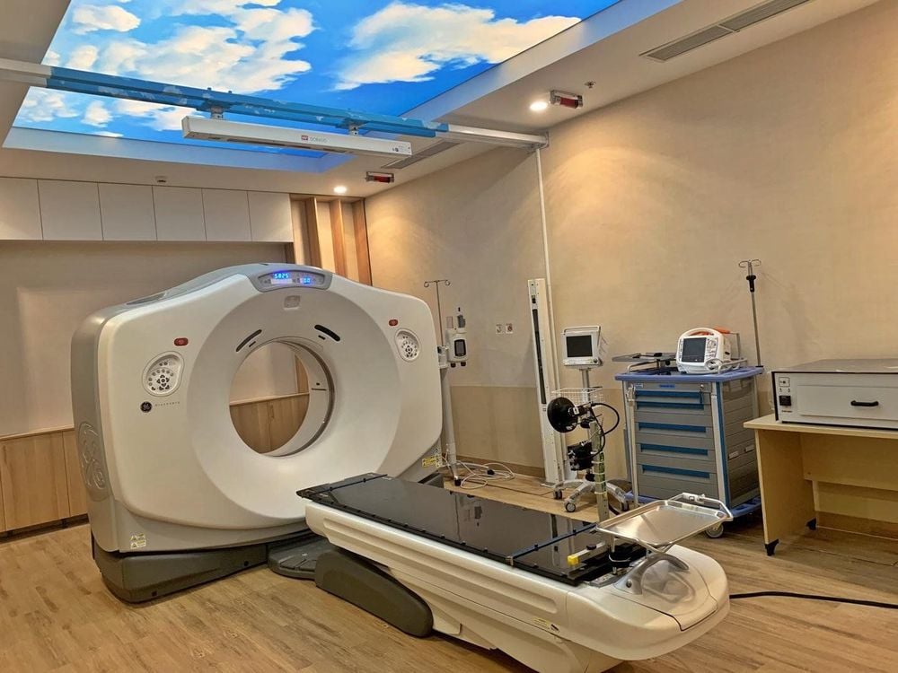
Simulated tomography is a very important step to help doctors plan and plan treatment accurately. Patients who have decided to perform a CT simulation need to follow the instructions of the doctor. Accordingly, to ensure the effectiveness, patients need to choose reputable medical facilities to perform this technique.
In order to meet the needs of medical examination and treatment, currently Vinmec International General Hospital has been and continues to bring in a system of modern machines such as magnetic resonance imaging (MRI), computed tomography (CT), X-ray, etc. CT simulation... in medical examination and treatment, diagnostic imaging, disease treatment. Accordingly, the imaging, diagnostic and follow-up procedures at Vinmec are all performed by experienced specialists, capable of early detection of many diseases since there are no symptoms. Especially in order to bring high efficiency in medical examination and treatment, Vinmec now also designs many accompanying medical services, bringing many conveniences to customers.
Please dial HOTLINE for more information or register for an appointment HERE. Download MyVinmec app to make appointments faster and to manage your bookings easily.


