This is an automatically translated article.
The article is expertly consulted by Dr., BS. Ton That Tri Dung - Head of Department of Examination & Internal Medicine - Department of Examination & Internal Medicine - Vinmec Da Nang International HospitalIncreased intracranial pressure is a syndrome that can cause dangerous complications such as cerebral edema, cerebral ischemia, even brain drop, causing irreversible damage or rapid death to the patient. Therefore, patients with signs of increased intracranial pressure need to be recognized early and treated promptly.
1. Signs of increased intracranial pressure
It is necessary to recognize signs of increased intracranial pressure through early and timely signs in order to quickly and properly handle it to limit the risk of life-threatening danger to the patient. Depending on the state of the patient, awake or unconscious, the patient will have different signs of increased intracranial pressure. Specifically:
In patients who are awake:
Increased intracranial pressure will cause signs of headache to increase gradually, the headache may be diffuse or localized Frequent vomiting of causes in the posterior fossa Disorder visual disturbances with signs of double vision, blurred vision, decreased visual acuity, ophthalmoscopy with papilledema Nervous disorders with lethargy, somnolence In comatose patients:
Patients with increased intracranial pressure are sudden awakening coma, or deeper coma Shows increased muscle tone Autonomic dysfunction - a serious sign of illness Fast or slow, irregular, raised blood pressure or possibly a drop in blood pressure Respiratory disturbance with rapid, deep, or Cheyne-Stockes breathing. Disturbance in body temperature regulation manifested as high fever or hypothermia. There are signs of brain damage caused by hypothermia such as temporal lobe drop, III cord paralysis, dilated pupils; Amygdala cerebellar hypoplasia presents with tachypnea or apnoea, central cerebral hypoplasia represents damage from top to bottom.

Tăng áp lực nội sọ ở bệnh nhân hôn mê
However, in most patients with increased intracranial pressure, there are general symptoms of increased intracranial pressure that are mentioned to quickly recognize this syndrome such as:
Headache: Headaches often occur all day The pain increases at night and in the morning, the location of the pain varies depending on the location of the lesion, depending on the cause, but can be pain in the forehead, temples, occipitals and eye area. Pain increases with exercise, exertion, cough and sneeze due to increased venous pressure, relieves pain when standing, sitting The patient may have nausea due to stimulation of the X cord, usually appearing after a headache, Vomiting easily in the morning, after vomiting will feel less headache. Dizziness is caused by frequent compression of the vestibular region or by damage to the vestibular center in the brain stem or in the temporal and frontal areas. Visual disturbances with diplopia, difficulty seeing due to VI nerve palsy, papilledema are the most objective symptoms of increased intracranial pressure, usually bilateral edema. Other typical signs of increased intracranial pressure such as:
Tinnitus Slow pulse, blood pressure changes tend to increase The initial breathing rate is normal after tachycardia, Cheyne - Stokes breathing There are signs of vegetative disorder such as: manifestations of sweating, cold extremities when severe abdominal pain, appearance of acute abdominal pain, constipation, increased temperature, vomiting black substance. Mental disorders often manifest as sluggishness, apathy, apathy, memory disturbances, dark consciousness, confusion, drowsiness or coma, even disorientation in space, time, sometimes Most schizophrenia are tumors in the temples. Especially in children under 5 years old, signs of increased intracranial pressure causing skull dilation are manifested by an increase in head circumference. The young skull has scalp varicose veins, the eyes are large, protruding, and the sound of "broken vase" can be heard as Macewen's sign. A murmur may be heard on the skull or in the eyes in cases of angiomas or vascular malformations.
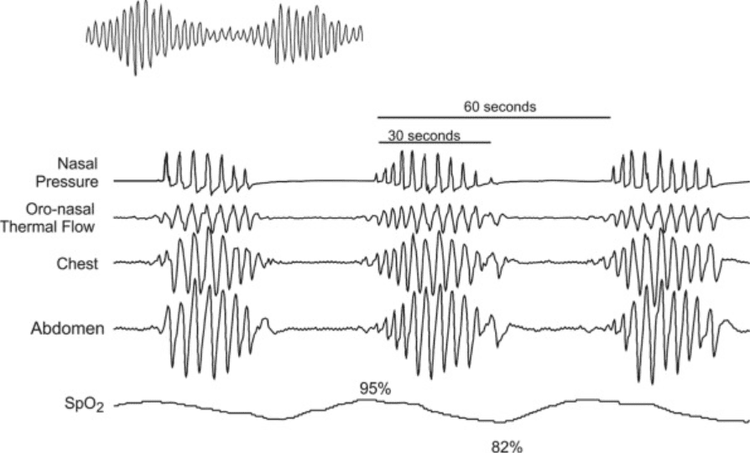
Thở Cheyne – Stokes
2. Subclinical signs of increased intracranial pressure
Blood tests can identify the cause of hyponatremia. Ophthalmoscopy: In the early stage, papilledema shows that the optic disc is fuller compared to the retinal surface and pinker than normal Stage of papilledema: The optic shoulder is completely erased, the optic nerve is swollen and swollen on the retina, as shown in Fig. mushrooms, red and pink thorns tend to grow out like flames. The blood vessels constricted. Hemorrhagic stage: There are more haemorrhagic clusters in the optic disc and retina Stage atrophy: In the decompensated stage, the optic nerve becomes white, losing its shine, and the edges are jagged. The blood vessels are sparse and pale in color, accompanied by clinically reduced patient's visual acuity. Cranial radiographs: Can see dilated skull joints, especially the top of the forehead in children, finger impressions pay attention to the temporal crest and fossa. Wide saddle but often late to show varicose veins on the skull, the cranial vault may be thin. EEG suggests localization, evaluates the severity of raised intracranial pressure. Transcranial Doppler ultrasound Computed tomography (CT-scan) of the brain: can see: Cerebral edema, brain structure is pushed, midline structure is changed. Ventricular dilation: Due to obstruction of the circulation of cerebrospinal fluid. Can see: Brain bleeding, cerebral ischemia, brain tumor, brain abscess... Magnetic resonance (MRI) of the brain: For more information about brain damage. Cerebral angiography: Identify cerebral vascular malformations. Lumbar puncture: when meningitis is suspected

Chọc dò tủy sống
Combining both the clinical and laboratory signs mentioned above, the patient can soon be accurately diagnosed with signs of increased intracranial pressure.
Vinmec International General Hospital with a system of facilities, fully equipped with modern machinery and medical equipment, the most advanced imaging equipment to support diagnosis and treatment such as: CT SCAN Toshiba 640 slices, cerebral angiogram, G.E 3.0 MRI (MRI) scanner, MRA and CTA... for clear image quality, easy diagnosis and treatment of brain and spine diseases of patients both medical and surgical pathology.... with a team of experts, doctors with many years of experience in the examination and treatment of neurological diseases, patients can completely rest assured to examine and treat neurological diseases. in general and intracranial pressure in particular at Vinmec.
To register for examination and treatment at Vinmec International General Hospital, you can contact Vinmec Health System nationwide, or register online HERE.


