This is an automatically translated article.
The article is expertly consulted by Master, Doctor Nguyen Hong Hai - Doctor of Radiology - Department of Diagnostic Imaging and Nuclear Medicine - Vinmec Times City International General Hospital.There are many imaging techniques used for ureteral stones such as ultrasound, X-ray, UIV... However, multi-slice tomography technique in evaluating ureteral stones is very useful for identifying ureteral stones. Stone characteristics, can evaluate most of the characteristics of ureteral stones according to clinical requirements, thereby making an important contribution in supporting the selection of treatment methods for ureteral stones.
1. Principle of multi-slice tomography machine
Multi-slice computed tomography scanner is a machine that increases the number of rows of X-ray detectors in the x-ray system, thereby increasing the number of images and the thinness obtained in the same unit of time.There are many types of multi-slice tomography machines with different rendering software advantages, which result in different image quality. The higher the scanning speed, the faster the capture time will be, the patient will not have to hold their breath many times or hold their breath for a long time when taking pictures. In particular, for patients who cannot lie still for a long time, it is necessary to choose a time when the patient lies still for a short time to be able to take a cranial sinus scan. Therefore, this is the advantage of this method.
In addition, the thinner the slice thickness, the closer to each other, the more images will be obtained. Therefore, the screening is more effective, not missing even the smallest lesions. At the same time, the images of the organs in the patient's body are also clearer and sharper.
2. Multi-slice tomography in the evaluation of ureteral stones
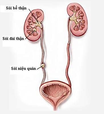
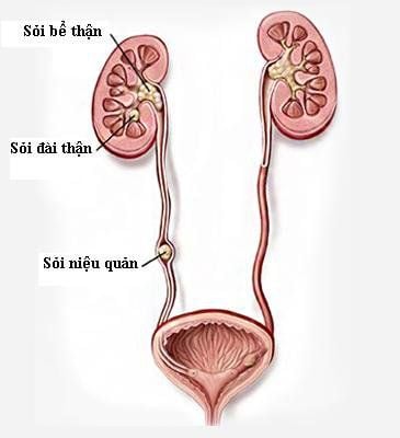
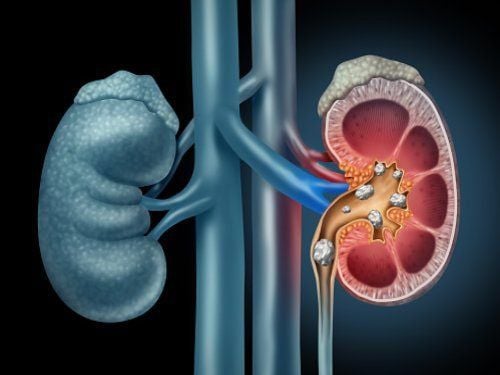
2.1. How dangerous are ureteral stones? If detected early, ureteral stones will have appropriate intervention to prevent the bad progression as well as dangerous complications of the disease. However, if people with ureteral stones do not detect the disease early or do not treat it in time, it can lead to some dangerous complications as follows:Obstruction of urine excretion: Causes of obstruction Urine is caused by stones located in the ureters, obstructing the flow of urine between the kidneys and bladder. Therefore, the upper urine is stagnant, causing the kidneys to retain water, compressing the renal parenchyma or causing the renal calyces to be dilated. Eventually, kidney function declines, leading to kidney failure. Bacterial infections: The urinary system is the place to contact excess waste products in the body that the body needs to excrete through urine. However, when stones form, the stones move will damage the lining of the ureters as well as other nearby organs, leading to urinary tract infections. If the infection is combined with hydronephrosis, it can cause pus in the kidney, sepsis and death. Kidney failure: This is the most dangerous complication for people with ureteral stones. This condition is usually caused by stones forming on either side of the urinary system or on one side of the kidney. 2.2. Meaning of multi-slice computed tomography in evaluating ureteral stones To diagnose ureteral stones, there are many imaging techniques used such as ultrasound, X-ray, UIV... However, the cutting technique Multi-slice layers in the evaluation of ureteral stones are useful for defining stone characteristics. Because multi-slice tomography technique can evaluate most of the features of ureteral stones according to clinical requirements, thereby making an important contribution in supporting the selection of treatment methods for ureteral stones.
In addition, the advantages of multi-slice tomography machine allow isotropic mass imaging, and polyhedron survey as well as algorithmic image processing to assist doctors in diagnosing stones. ureter, and accurately assess the number, location and size of stones. In addition, the multi-slice tomography technique in evaluating ureteral stones also supports the doctor in:
Assessing whether the ureter is watery: Reconstructed image of the frontal plane with high resolution of the scanner. Multi-slice tomography allows rapid but very accurate assessment of ureteral stones. At the same time, the multi-slice tomography method can also determine the fragility and composition of stones thanks to the density and characteristics of the stone structure. Finding signs of ureteral stones: Most ureteral stones are detected on multi-slice tomography without intravenous contrast. At the same time, multi-slice computed tomography can also help identify indirect signs such as pyelonephritis, perirenal adipose tissue edema, ureteral dilatation on stones, edema of periureteral fatty tissue, kidney with stones. enlarged, from which there is an early intervention method because the possibility of stone drainage is extremely low. Determination of stone composition: The composition of ureteral stones is considered one of the most important factors in choosing the appropriate treatment for the patient. Stone density on multi-slice tomography helps predict stone composition. If the multi-slice tomography image shows uric acid stones, the treatment of urinary alkalinization is the first priority method. Similarly, if the stones are calcium or cysteine-based, it is difficult to break up with extracorporeal shock wave lithotripsy. Multi-slice tomography is used to monitor after treatment of ureteral stones: The main purpose of multi-slice tomography to examine images after treatment of ureteral stones is to determine the status of stones (clearance of stones). or not), rule out urinary tract obstruction as well as residual stones. At the same time, multi-slice tomography also supports the detection of complications after treatment interventions such as hematomas or urinary cysts.
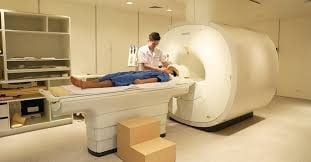
Patients need to go to a reputable hospital to conduct examination and treatment as soon as there are signs of ureteral stones. Currently, Vinmec International General Hospital is one of the leading prestigious hospitals in the country, trusted by a large number of patients for medical examination and treatment. Not only the physical system, modern equipment: 6 ultrasound rooms, 4 DR X-ray rooms (1 full-axis machine, 1 light machine, 1 general machine and 1 mammography machine) , 2 DR mobile X-ray machines, 2 multi-row computer tomography rooms with receivers (1 256-series machine, 1 512-series machine and 1 16-series machine), 4 Magnetic resonance imaging rooms (3 3 Tesla machines and 1 1.5 Tesla machine), 1 room for interventional angiography with 2 aspects and 1 room for measuring bone mineral density.... Vinmec is also the place to gather a team of experienced doctors and nurses who will assist a lot in the treatment. Diagnosis and early detection of abnormal signs of the patient's body. In particular, with a space designed according to 5-star hotel standards, Vinmec ensures to bring the patient the most comfort, friendliness and peace of mind.
Please dial HOTLINE for more information or register for an appointment HERE. Download MyVinmec app to make appointments faster and to manage your bookings easily.


