This is an automatically translated article.
Laparoscopic surgery to open the common bile duct to remove stones is a method of treating common bile duct stones with many advantages such as small scars, less pain, early mobilization, reduced rate of complications after surgery, ...
1. Treatment of common bile duct stones
Gallstone is a very common surgical disease in Vietnam. In particular, common bile duct stones account for about 50-60% of all cases of gallstones. In European and American countries, common bile duct stones are often secondary to gallstones moving down, most commonly cholesterol stones due to metabolic disorders. In Asian countries, including Vietnam, the main causes of common bile duct stones are parasites and bacterial infections. Stone components are usually bile pigments made from eggs or carcasses of roundworms.
When newly infected with common bile duct stones, patients often have no obvious symptoms, the main signs are flatulence, abdominal distension, indigestion. When the stone grows to a large size, causing obstruction of the biliary tract, it will cause prolonged severe abdominal pain, pain may spread to the shoulder or back; high fever of 39-40 degrees occurs at the same time or several hours after the pain; Jaundice of the skin and mucous membranes of the eyes.
Common bile duct stones, if not detected and treated promptly, can lead to dangerous complications such as gallbladder rupture, perforation of the common bile duct causing peritoneal infection; pyelonephritis, acute pancreatitis due to gallstones, acute gallstone nephritis (hepatobiliary syndrome), septic shock of the biliary tract,... These are all serious conditions, complicated treatment, patients at risk chance of death.

Sỏi ống mật chủ không điều trị kịp thời sẽ gây ra nhiều biến chứng nguy hiểm
Stone removal surgery is the main method in the treatment of common bile duct stones. Before surgery, patients are usually treated medically with antibiotics, choleretics, biliary smooth muscle relaxants, intravenous fluids to provide water and electrolytes, etc. Patients are treated medically. , surgery often achieves better results, less complications after surgery. There are many surgical ways to treat common bile duct stones such as: endoscopic retrograde cholangiopancreatography (ERCP), open surgery, laparoscopic surgery, .. In which, laparoscopic open choledocholithiasis has many advantages such as scarring. Small, less invasive, so the patient has less pain after surgery, can move early, the ability to stick to the intestines is significantly reduced compared to open surgery,... Due to many advantages, laparoscopic surgery opens the common bile duct. Stone removal is done more and more widely.
2. How is laparoscopic open bile duct stone removal performed?
2.1. Indications for laparoscopic choledochalotomy to remove stones The basic content of laparoscopic cholecystectomy to remove stones is to access the intra-abdominal components through laparoscopy, perform dissection, reveal, Dissect the common bile duct or common hepatic duct to remove bile duct stones, then suture the biliary incision. Laparoscopic surgery to open the common bile duct to remove stones is indicated in the following cases:
Extrahepatic bile duct stones, with no limit on the size and number of stones. Extrahepatic biliary tract dilated >1cm. Laparoscopic surgery to remove stones is not performed when the patient has contraindications to laparoscopic surgery such as a history of laparotomy, inability to pump CO2 into the abdominal cavity such as heart failure, respiratory disease, ... In addition, patients with severe coagulopathy or respiratory and cardiovascular diseases that do not allow general anesthesia are also not allowed to perform this surgery.
2.2. Preparing the patient before surgery Before surgery, the patient will be performed basic tests such as biochemistry, hematology, liver function test and respiratory function. In addition, imaging techniques such as abdominal ultrasound, magnetic resonance imaging or computed tomography will be performed to survey the size of the extrahepatic bile ducts and diagnose gallstones.

Người bệnh tiến hành các xét nghiệm cần thiết trước khi phẫu thuật
2.3. Steps to conduct laparoscopic open bile duct stone removal The patient is instructed to lie supine on the operating table, legs closed or 90 degrees, left arm closed, right arm closed or flexed. The patient was then given endotracheal anesthesia and a nasogastric tube inserted. If the primary surgeon stands between the patient's legs, the monitor is placed on the right side of the patient's head. If the primary surgeon stands on the left side of the patient, the monitor is placed on the right side of the patient at hip level. The doctor placed the 10mm trocar just below the navel. Then, inflate CO2 into the abdomen, maintain intra-abdominal pressure 10-12mmHg, put the camera into the abdomen. Place the next 3 trocars, including: 1 10mm trocar and 2 5mm trocars, the location of these trocars is usually below the sternal tip, under the right and left ribs. Open the peritoneum covering the common bile duct, exposing the surrounding open tissue to see the anterior wall of the common bile duct. Use an electrocautery knife or scissors to dissect the common bile duct. Then, using pliers to pick up the stone, put it through the epigastric trocar hole to remove the stone in the bile duct, and at the same time pass it through Oddi into the duodenum. The plastic tube is inserted through the epigastric trocar into the common bile duct to flush the bile ducts, remove small stones, biliary pus, blood clots and check the communication of the end of the common bile duct and the sphincter of Oddi. If there is an intraoperative cholangioscope, the lens will be passed through the epigastric trocar, cholangioscopy to remove stones, check for stones and the state of communication of the end of the common bile duct and the sphincter of Oddi. Depending on the case, the doctor will put a drain or not. Suture the common bile duct with slow dissolving sutures. Aspiration washed, placed a subhepatic drain through the trocar foramen below the ribs. If Kehr drainage is present, insert Kehr through the epigastric trocar foramen. Gauze and gravel in a plastic bag are taken through the umbilical trocar hole. The trocar holes are closed with melt sutures. After surgery, the patient will have a nasogastric tube removed and early feeding. The amount and nature of the patient's drainage fluid will be monitored daily. The time to remove the Kert tube is usually 10-14 days if the patient's condition is stable, there are no complications, no abdominal pain, and no fever.
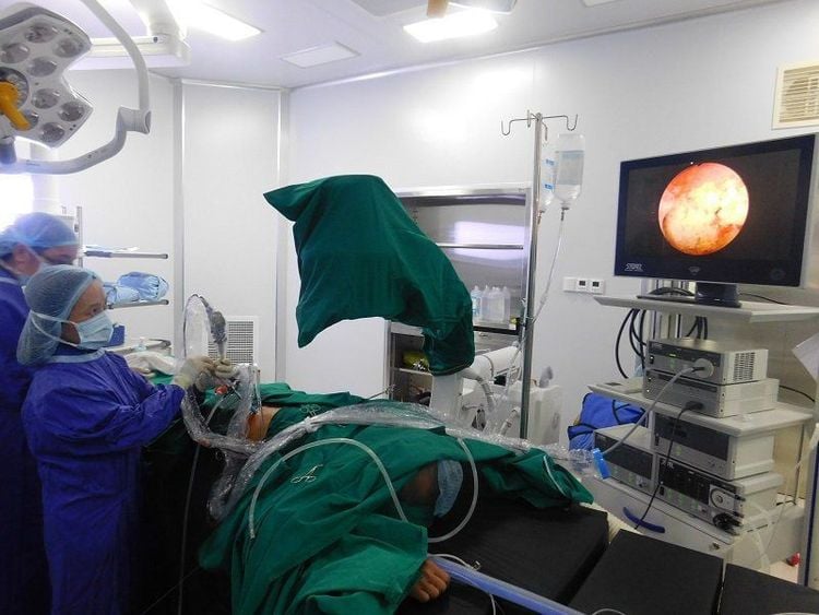
Phẫu thuật nội soi mở ống mật chủ lấy sỏi
3. Possible complications in laparoscopic open bile duct stone removal surgery
A common complication during surgery is abdominal bleeding, depending on the degree of bleeding, hemostasis is carried out laparoscopically or converted to open surgery if necessary.
Common postoperative complications are:
Residual abscess: The patient will be treated with antibiotics combined with aspiration under ultrasound. If the treatment is not successful, it will be switched to open surgery for treatment. Peritonitis : May be due to perforation of the common bile duct suture, primary biliary tract injury, or gastrointestinal tract injury. Patients are often re-operated to repair the lesion.
Please dial HOTLINE for more information or register for an appointment HERE. Download MyVinmec app to make appointments faster and to manage your bookings easily.


