This is an automatically translated article.
The article is professionally consulted by Specialist Doctor I Nguyen Hung - Department of Medical Examination & Internal Medicine - Vinmec Danang International General Hospital.
Ureteral stones are rarer than kidney stones. Although ureteral stones are easy to identify, they affect kidney function at an early stage. Let's find out what ureteral stones are as well as the characteristics of the disease in the article below.
1. What are ureteral stones?
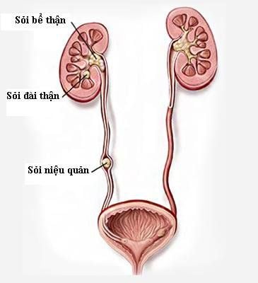
Ureteral calculi is a disease of the urinary tract where stones form in the kidney and travel down the ureter, where they end up in the natural narrowing of the ureter. Most ureteral stones have a width less than 5mm compared to the ureter, will follow the urine stream down the ureter naturally.There are two types of ureteral stones, which are:
Body stones (primary stones): Due to a biochemical disorder in the body; Organ stones (secondary stones): Due to blocked excretory tract leading to urine stagnation causing stones.
2. Progression of ureteral stones
The passage of stones from the kidney to the bladder depends on the size and location of the stone. Distal ureteral stones will travel down the bladder more easily than mid and proximal ureteral stones. However, the width of the stone is the main factor affecting stone movement; The passage of ureteral stones into the bladder is about 4-6 weeks. For stones with a size of 2 - 4 mm, the stones go down the bladder in 31 - 40 days; The rate of stones moving spontaneously according to the location of the stone was 22% (in the proximal ureter), 46% (in the middle ureter) and 71% (in the distal ureter); Depending on the location of the stone, the medical method of waiting for the stone to move naturally will be applied to ureteral stones less than 5mm in size. In contrast, with stones larger than 5mm, it is very difficult to move naturally. At this time, it is necessary to consider interventional treatment methods to remove stones; The locations of stones prone to entrapment are at the junction of the renal pelvis and ureters (10%), the place where the ureters cross the iliac vessels (20%), the intramural ureteral segment of the bladder (70%); When moving, ureteral stones cause damage to the ureters. The ureter around the stone is edematous, allowing the stone to stick to the ureter and not be able to move.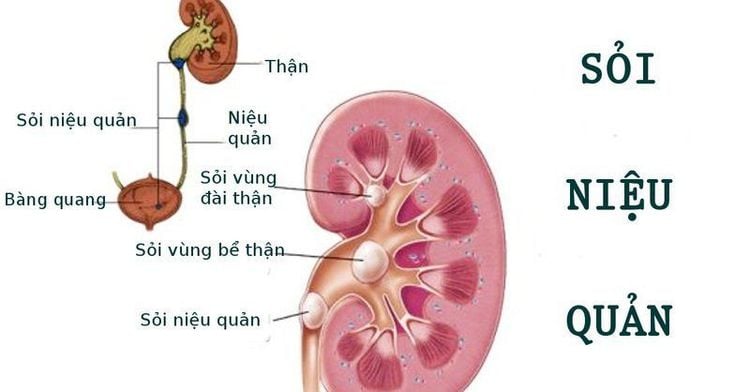
3. Clinical symptoms of ureteral stones
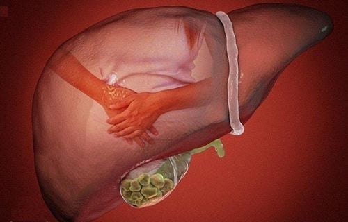
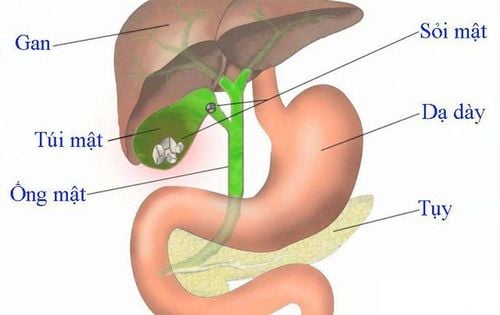
Ureteral stones cause renal colic as the stone moves through the ureter and causes ureteral spasm, the pain often comes on suddenly. Ureteral stones often cause nephrotic pain because the stones are mobile due to ureteral peristalsis and the relatively small size of the stones; The ureter has nerves, so when the stone moves and rubs against the mucosa, it will cause a constriction response; Urination, painful urination due to irritation of the bladder lining, painful ejaculation when the seminal vesicles and vas deferens are inflamed, possibly due to ureteral stones in the pelvic region, near the bladder; Blood in urine with micro or macroscopic appearance; Cloudy and purulent urine: This is a sign of reverse kidney infection, when fever is accompanied by chills. This situation can seriously threaten kidney function, there is a risk of sepsis and septic shock; Acute anuria is caused by ureteral stones causing urinary tract obstruction. However, this is a sudden symptom, when the urinary obstruction is resolved, the kidney will be restored; Chronic kidney failure with symptoms related to the digestive system such as bloating, flatulence, vomiting, indigestion, anemia, insomnia, etc., is caused by ureteral stones on one or both sides for a long time. that go undetected and untreated; Back pain, especially when working or doing heavy work, because the stone is not mobile and does not obstruct the urinary tract for a long time. Other clinical symptoms of ureteral stones can cause confusion with other diseases such as cholecystitis, duodenal ulcer, gastritis (right stone), descending colon (left stone). ..
Positive kidney touch if the kidneys are waterlogged, pain in the back and hips if inflammation around the kidney; Pain on pelvic examination, this may be a sign of middle ureteral stones, especially when there is an inflammatory reaction around the ureter; Pain under the ribs but without abdominal cramping, may be a sign of an upper ureteral stone.
4. Diagnosis of ureteral stones based on subclinical symptoms
Urinalysis and urinalysis show polycythemia vera, possibly white blood cells and bacteria. An infection of the urinary tract can cause albumin in the urine; X-ray of the unprepared urinary tract (KUB) helps to determine the location, size and shape of the stone, thereby allowing to predict the natural elimination capacity of the stone and the treatment regimen; Abdominal ultrasound helps to detect stones and hydronephrosis. Ultrasound also allows visualization of ureteral stones, especially those with low contrast. However, ultrasonography cannot describe the location of the stone and the function of the kidney; Contrast X-ray of the urinary system (UIV) is applied to ureteral stones requiring surgical intervention. X-rays allow determining the location of the stone in the urinary tract and the degree of dilatation of the pyelonephritis. In addition, this technique also reflects the function of the kidney with stones and the remaining kidney. In cases of severe hydronephrosis, it may not be excreted on UIV, therefore, imaging should be done 2, 4, or 8 hours after contrast injection. If the kidneys still do not excrete it is likely that the kidneys have been severely damaged. In case of stones in both kidneys, UIV is applied to select the preferred kidney for surgery. In addition, UIV also reflects the degree of dilation of the ureter above the stone; Ureterography - retrograde pyelonephritis (UPR) is applied when the above techniques have not yet concluded whether the patient has ureteral stones or not. However, this is an invasive procedure and can introduce bacteria from the urethra to the upper urinary tract. Currently, this technique has been replaced by CT scanning; CT scans can identify ureteral stones and the degree of obstruction, as well as assess kidney function. Dr. Nguyen Hung graduated as a doctor specializing in General Internal Medicine - University of Medicine and Pharmacy, Ho Chi Minh City; Can speak English and French. With over 36 years of experience in the profession, of which 17 years is the Head of the Department of Endocrinology - Endocrinology, Da Nang Hospital, the doctor has experience in treating endocrine - diabetes and kidney diseases. Currently, he is an endocrinologist at the Department of Medical Examination & Internal Medicine at Vinmec Danang International General Hospital.Ureteral stone is a disease that causes renal colic pain, the diagnosis is mainly based on X-ray method. Patients with suspected symptoms of ureteral stones should see a doctor for the most accurate diagnosis.
Please dial HOTLINE for more information or register for an appointment HERE. Download MyVinmec app to make appointments faster and to manage your bookings easily.


