This is an automatically translated article.
The article is expertly consulted by Master, Doctor Nguyen Hong Hai - Doctor of Radiology - Department of Diagnostic Imaging and Nuclear Medicine - Vinmec Times City International General Hospital.Along with semen analysis, scrotal ultrasound is an effective way to diagnose abnormalities of the testes, epididymis and spermatic cord causing infertility in men. Scrotal ultrasound is simple and non-invasive, helping to accurately diagnose diseases in the scrotum.
1. Anatomy
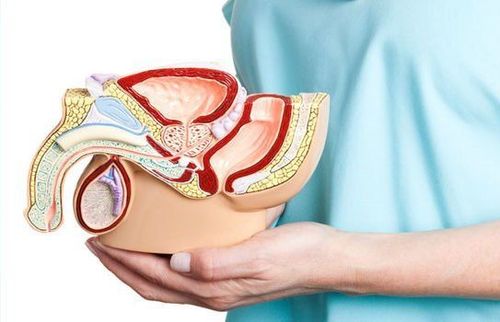
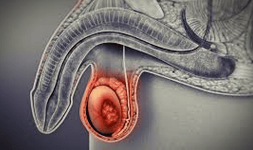
The scrotum is the male sex organ located under the penis and contains the testicles, seminal vas deferens, spermatic cord, and epididymis. A thin membrane of the scrotum lining the scrotal cavity and organs, which adhere closely to the fibrous layer covering the testicle.Testicles are bean-shaped, about 4x3x2.5cm, weighing about 20g. Testicles have endocrine and exocrine functions.
The epididymis is about 6cm long, 0.6 - 1.5cm thick, covering the testicle posteriorly. The tail of the epididymis continues to form the vas deferens, which run upward out of the scrotum. The spermatic cord contains arteries, veins, vas deferens, and veins of the venous plexus that run up the inguinal canal into the abdomen.

2. How is a scrotal ultrasound performed?
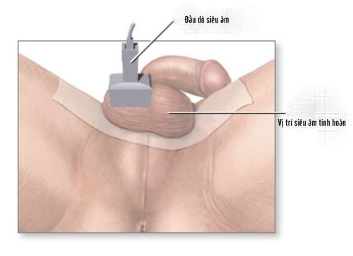
The patient lies supine, legs slightly apart, holding the penis and pulling it slightly toward the head to expose the scrotum. Ultrasound uses a flat high-frequency transducer that scans the transducer with planes along the longitudinal and transverse axes. Regular ultrasound of both testicles to compare, contrast and analyze anatomical signs.Ultrasonography of the scrotum will thoroughly probe for abnormal acoustic structures, examining the epididymis and spermatic cord. Doppler ultrasonography for low-velocity vascular flow exploration is of high value in the diagnosis of epididymitis, testicular torsion, and varicocele. Usually performed together with energy Doppler and spectral Doppler, a new comprehensive ultrasound helps evaluate the entire urinary system.
3. What do scrotal ultrasound results show?
Comprehensive ultrasound imaging helps the doctor assess the morphology, size of the scrotum and male genitals.3.1. Normal ultrasound results
When the testicles are normal, the parenchyma is even. Occasionally, blood vessels can be seen. Testicular margins are thin, membranous, hyperechoic lines. The umbilicus is a thin, hyperechoic panel inside the testicle. The parietal and visceral leaves are dark and light lines. Ultrasonography is difficult to detect testicular appendages and epididymis. The epididymis has a similar acoustic structure and is slightly lighter than that of the testis. Body and tail slender, slightly increased sound. The head and tail are clearly visible, and the physiological fluid layer can be clearly seen. On Doppler ultrasound, it is easy to see the seminal artery, with a curve corresponding to the parenchymal artery. Veins are low-velocity flow. The vine is an image of structures with multiple hypoechoic lines in the longitudinal section and a circular structure with hypoechoic circles in the cross section.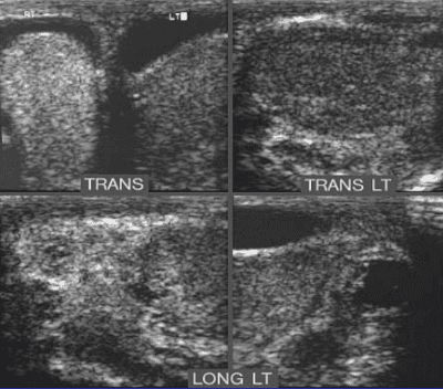
3.2. Pathological ultrasound results
Abnormal scrotal ultrasound image due to pathology shows:Image of testicles increasing, decreasing in size. Homophonic reduction, boost, mixed sound. See dilated vascular structure in varicocele. Localized lesions: cysts, calcifications, abscesses, tumors.... Diffuse lesions: inflammation, diffuse microcalcification,...
4. Common pathology and ultrasound results
Most common diseases in the scrotum can be detected by ultrasound.4.1. Congenital pathology
Ectopic testicle: The scrotum is small, often hypoechoic in the inguinal canal and pelvic fossa. Vulnerable to injuries, twists. Testicular atrophy. Multiple testicles.
4.2. Inflammatory disease
Orchitis, epididymitis Subacute epididymitisPatients with subacute epididymitis under 40 years old are usually caused by Chlamydia Trachomatis, over 40 years old are often due to lower urinary tract diseases. The epididymis is large, if bleeding occurs, hyperechoic spots appear. The disease can cause infertility complications, epididymal fibrosis, abscesses, testicular atrophy, testicular infarction. Acute orchitis
Large size, echocardiography shows diffuse reduction, possibly necrosis. Chronic orchitis
The picture is heterogeneous, showing fibrosis, thick tunica, possibly calcified nodules. Epididymitis has little effect, has 2 forms:
Diffuse orchitis: diffuse interstitial fibrosis, rarely seen on ultrasound.
Scrotal stones Visible calcification nodules.
Tuberculosis Sees forming an abscess, the calcification is getting bigger and bigger, there may be fluid. Tuberculosis usually affects the epididymis first, then the testicles.

Tumors outside the testicles: Wall of the scrotum, epididymis; Benign testicular tumor; Secondary malignancies.
Hematocele: large multiseptal and echogenic fluid collections compress the contents of the scrotum.
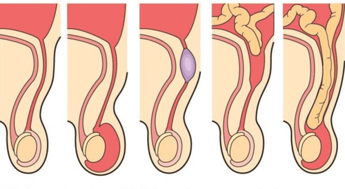
In addition, ultrasound also detects abnormal scrotal images due to other diseases such as scrotal hernia, testicular cyst, epididymal cyst, spermatic cord cyst,...
Thus, scrotal ultrasound plays an important role It is important in the diagnosis, detection and treatment of diseases related to the genital organs, the most common of which is male infertility. The results of a scrotal ultrasound may need to be combined with other evaluation tests to make an accurate diagnosis.
When the male genitals appear abnormal symptoms, you should go to a reputable medical facility for timely examination and treatment. Vinmec International General Hospital is a high-quality medical facility in Vietnam. Modern, state-of-the-art medical equipment imported from the US, Japan, the Netherlands... helps diagnose and treat quickly and effectively. The doctors are highly qualified, experienced, and trained abroad.
Master, Doctor Nguyen Hong Hai graduated with a Master's degree in Diagnostic Imaging at Hanoi Medical University with strengths in diagnosing breast and thyroid cancer. Currently, Dr. Hai is working at the Department of Diagnostic Imaging, Vinmec Times City International Hospital.
Please dial HOTLINE for more information or register for an appointment HERE. Download MyVinmec app to make appointments faster and to manage your bookings easily.


