This is an automatically translated article.
Radiation in medicine (mainly emitted from equipment such as X-ray machines, radiotherapy machines) and can cause some adverse problems to the health of patients and doctors.
1. What is radiation in medicine?
Ionizing ray radiation includes:
High-energy electromagnetic waves: gamma rays, X-rays Types of particles: alpha particles, beta particles and neutrons. In medicine, ionizing radiation is mainly emitted from radioactive elements and devices such as X-ray machines and radiation therapy devices.
Most imaging modalities that use ionizing radiation (X-rays, CT scans or scintigraphy) expose patients to relatively low doses of radiation, which are generally considered safe. . However, all types of ionizing radiation have the potential to be harmful to health. There is currently no safe threshold that can completely rule out adverse effects. Therefore, it is necessary to focus efforts to bring the level of radiation exposure in medicine to a minimum.
How to measure radiation exposure:
Absorbed dose: The amount of radiation absorbed per unit mass, with units of gray (Gy) and milligray (mGy). Previously, it used radiation absorbed dose unit (rad): 1mGy - 0.1 rad. Equivalent dose: The absorbed dose times the radiation weight factor adjusted for effects on tissues, based on the type of radiation. The units used are sieverts (Sv) and millisieverts (mSv). Previously, it used the human roentgen equivalent magnetic field unit (rem; 1 mSv = 0.1 rem). For X-ray imaging facilities, the radiation weight factor is 1. Effective dose: One of the measures of cancer risk. It helps to adjust the dose equivalently based on the susceptibility of the exposed tissues (eg the genital system is the most vulnerable organ). Use Sv and mSv units. The effective dose will be higher in younger subjects.
2. Radiation risk in medicine
Medical radiation can be harmful if the total cumulative dose is high. Examples include multiple CT scans (CT scans require a higher dose of radiation than most other imaging modalities). In addition, radiation exposure in medicine also needs attention in some high-risk cases such as: pregnant women, infants, children 1 - 4 years old, young women needing mammograms to diagnose cancer breast .
2.1 Radiation and Cancer Scientists estimate there is a cancer risk from radiation exposure in diagnostic imaging. Although this risk is relatively low, it still exists if the radiation dose used reaches the threshold of tens of mGy (equivalent to the radiation threshold used in CT scans). The CT pulmonary angiogram (to detect pulmonary embolism) delivers a radiation dose to the breasts that is about 10-25 times higher than that of a two-position mammogram.
Young patients have a high risk of cancer due to radiation in medicine because:
Life is longer, so cancer has more time to develop; The cell growth rate is stronger than in the elderly, so it is more sensitive to DNA damage.

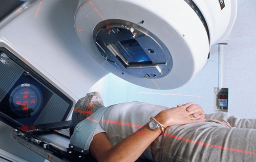
Nhiễm xạ trong y khoa người bệnh có nguy cơ ung thư do phơi nhiễm bức xạ
In addition, the risk of cancer depends on which tissue is irradiated. Lymphatic tissues, blood, bone marrow, ovaries, testes, and intestines are all very sensitive to radiation. In adults, the musculoskeletal system and the central nervous system are relatively resistant to radiation.
2.2 Radiation in Pregnancy The radiation risk depends on: The dose, what the imaging tool is, and what body area the diagnostic scan is being performed on. The fetus will be exposed to less radiation than the mother. In addition, the fetus will be exposed to negligible radiation if the mother: X-ray of the head, cervical spine, extremities and mammogram while the uterus has been screened.
In addition, the radiation exposure of the uterus depends on factors such as gestational age and the size of the uterus. The effects of radiation will depend on gestational age (from the time of conception).
3. Manifestations of radiation exposure
When infected with radiation, patients often have the following symptoms:
Mild form: Nervous system disorder, tachycardia, low arterial blood pressure, sinus arrhythmia, intestinal motility disorder and biliary function, easy stimulated Progressive form: Clinical and electrocardiographic manifestations of myocardial dystrophy, persistent hypotension, persistent bone marrow hypoplasia, thrombocytopenia, ovarian dysfunction, female less menstruation ; Chronic radiation dermatitis: Symptoms include pain, itching, dry skin, dysesthesia, nail dystrophy, hyperkeratosis, congestion, skin ulceration, cracking,...; Late signs: Bone cancer, skin cancer, myeloid leukemia, lung epidermal cancer,...

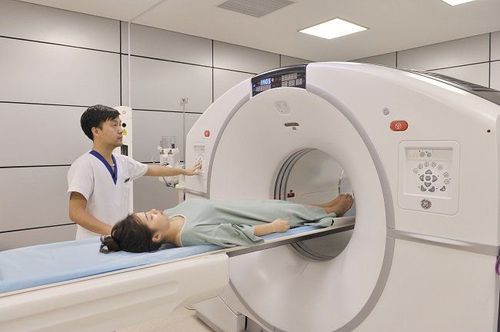
Tia xạ trong y khoa có thể khiến người bệnh bị nhiễm xạ với các thể khác nhau
4. Measures to prevent radiation contamination in medicine
In the US, CT scans account for only 15% of total scans, but this technique accounts for up to 70% of total radiation emitted by all imaging facilities. Compared with single-sequence CT, multi-sequence CT has 40-70% more radiation. However, recent advances in science and technology such as automatic exposure control or repeat reconstruction algorithms and 3rd generation CT receiver arrays,... have contributed to a significant reduction in dose. radiation used in each CT scan.
Faced with concerns about the increased risk of radiation exposure when performing diagnostic imaging techniques, the American College of Radiology has initiated the following programs: Image Wisely (for adults) and Image Gently (for children). These are programs that provide resources and information on minimizing radiation exposure for radiologists, clinicians, other medical professionals, and patients.
To reduce the risk of medical radiation exposure, imaging facilities with ionizing radiation should be indicated only when necessary. Physicians should consider alternatives. For example, in young children, if there is a minor head injury, diagnosis and treatment can be based on clinical evidence. Appendicitis can be diagnosed by ultrasound. However, if the potential benefits outweigh the potential risks, doctors should not delay, if necessary, to take a scan immediately, even with high radiation doses such as performing a CT scan.
Before carrying out the imaging facilities with ionizing radiation on women of childbearing age, it is necessary to pay attention to the issue of the fetus (the risk of radiation exposure is highest in the first 3 months - the period of pregnancy). where few people realize they are pregnant). If diagnostic imaging is necessary, care should be taken to shield the uterus for the above subjects (if possible).
In addition, occupational radiation sickness has no specific treatment, but needs comprehensive treatment, adequate rest, adequate nutrition (protein, vitamins B12, B6) and anti-diarrheal drugs. blood, blood transfusion, ... For prevention, should stay away from radioactive sources when handling, have walls and screens suitable for different types of radiation. In addition, attention should be paid to the recommended safe time for each type of radiation.
Medical staff when working or handling radioactive substances need to wear protective clothing, other protective equipment, lead plates, leaded rubber gloves, waterproof clothing, shower wash immediately after working hours (helps protect against irradiation, internal irradiation and against irradiation). At the same time, it is necessary to have regular health checks every 3-6 months, paying attention to blood tests to detect early signs of radiation-related diseases. In particular, attention should be paid to chronic skin lesions (if frequently exposed to radiation sources).
Medical radiation can enter the body through the skin, the digestive tract or the breath. Doctors and patients need to absolutely follow safety regulations to minimize the risk of radiation exposure.
Please dial HOTLINE for more information or register for an appointment HERE. Download MyVinmec app to make appointments faster and to manage your bookings easily.


