This is an automatically translated article.
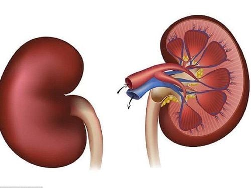
The article is professionally consulted by Master, Doctor Le Thi Minh Huong - Doctor of Resuscitation - Emergency, Department of Resuscitation - Emergency, Vinmec Nha Trang International General HospitalThe location of the renal artery is retroperitoneal, giving rise to several small branches before dividing into terminal branches into the renal hilum. The function of the renal artery is to provide the renal parenchyma with oxygenated blood for nourishment. It also provides blood for filtration, removing metabolic wastes and excess water.
1. Renal Vascular Anatomy
The renal arteries branch from the abdominal aorta and supply blood to the kidneys. The renal artery supply is variable from person to person and there may be one or more renal arteries supplying blood to each kidney. Due to the location of the aorta, inferior vena cava as well as renal anatomy in the body, the right renal artery is normally longer than the left renal artery.The blood supply to the kidney begins with the bifurcation of the aorta into the renal arteries (each named after the region of the kidney they pass through) and ends with the continuation of the renal veins to connected to the inferior vena cava. The renal arteries divide into several segmental arteries as they enter the kidney, which then divide into many arterioles.
The afferent renal arterioles branch into the glomerular capillaries, facilitating blood supply to the nephrons within the Bowman's capsule. Simultaneously, blood is taken out of the glomerulus and into the streptococcal capillaries, providing tissue oxygen to the renal parenchyma.
The kidneys of a normal adult receive about a quarter of the blood supply from the heart or 1.2 liters of blood per minute. The body has self-regulatory mechanisms in place, increasing or decreasing blood flow in response to blood pressure in the circulatory system. The receptors are located in the smooth muscle wall of the renal artery that allows the artery to dilate or contract to regulate the balance for high or low blood pressure.
2. Anatomy position and relationship of renal artery
The renal artery arises perpendicular to the abdominal aorta just below the branch of the superior mesenteric artery, roughly level with the transverse disc between the L1 and L2 vertebrae. Here, each side of the inferior aorta divides into the right renal artery and the left renal artery that supplies blood to the ipsilateral kidney.Anatomy of the right renal vein: Anteriorly, the proximal part of the right renal artery is related to the inferior vena cava, the distal part of the right renal vein is shorter. These structures separate the second part of the duodenum and the first part of the pancreas. Posteriorly, the right renal artery is associated with the right renal pelvis and distal ureter. Anatomy of the left renal vessel: The left renal vein separates the left renal artery from the body and tail of the pancreas and the splenic vessel. Posteriorly, the proximal part of the left renal artery is related to the left diaphragm, the left main psoas muscle, the left sympathetic trunk, and the body of the 2nd lumbar vertebra. The distal part of the left renal artery is related to the cistern. The left kidney and ureter are located posteriorly.

Giải phẫu mạch thận có vị trí bắt nguồn từ động mạch chủ bụng và đi vào ống thận nhằm cung cấp máu cho thận
3. Anatomy of the bifurcations of the renal artery
Before entering the renal hilum to supply blood to the kidney, the renal artery on each side divides into several branches for neighboring organs, such as the adrenal gland, the ureter, the perirenal mass, the renal capsule, and the renal pelvis. The renal artery then terminates near the renal hilum by dividing into anterior and posterior branches:Posterior branch : Passes posteriorly to the renal pelvis and supplies the posterior region of the kidney. Anterior branch: Subdivides further into segmental arteries, which supply renal segments, giving rise to apical, superior, anterior inferior, and inferior segmental arteries that each supply respective segments of they. Next, the segmental arteries divide into lobar branches, usually one for each renal pyramid. When reaching the small calyces, they divide again into interbranching arteries, which become arcuate arteries at the base of each calyx. The arcuate arteries then enter the nephrons as interstitial arteries and finally terminate in the glomerulus.
4. Anatomical variations of renal vessels
Differences in the number, origin, and path of renal arteries are quite common.Single renal artery: Normally occurs about 70% of cases, while accessory renal artery has about 30% of cases. The accessory renal artery usually arises just below or in some cases above the main renal artery. Rarely, the accessory renal artery arises from the visceral trunk or the superior mesenteric artery. The origin of the renal artery may vary, with one or both renal arteries arising from the bifurcation of the aorta or from the common iliac.
In summary, the renal anatomy is located at the origin of the abdominal aorta and into the renal tubule to supply blood to the kidney. Any variation in the blood supply to the 2 kidneys is important to clinicians prior to surgery or other invasive renal procedures on the kidneys.
Please dial HOTLINE for more information or register for an appointment HERE. Download MyVinmec app to make appointments faster and to manage your bookings easily.