This is an automatically translated article.
The article is made by Master, Doctor Ton Nu Tra My - Department of Diagnostic Imaging - Vinmec Central Park International General Hospital.
Prostate magnetic resonance imaging is a method used to diagnose suspected prostate cancer or follow up after treatment. Prostate MRI is performed according to a standard procedure to obtain an accurate diagnosis.
1. An overview of prostate cancer
The prostate gland is located below the bladder and above the rectum, next to the seminal vesicles. The size of the prostate changes, increasing with age. Prostate cancer is a condition in which cells in the prostate gland grow abnormally and become malignant. Most prostate cancer cells are of the adenocarcinoma type. Usually, prostate cancer grows very slowly, without symptoms in the early stages, so many patients are only detected when the disease has progressed to a late stage, leading to many difficulties in treatment and risks. high mortality.
Factors that increase the risk of prostate cancer include: Age (people aged 50 - 65 years old), family factors, unhealthy diet, being overweight or obese, smoking. , prostatitis , sexually transmitted disease , vasectomy , chemical exposure,...

Thừa cân béo phí là một trong các yếu tố làm tăng nguy cơ ung thư tuyến tiền liệt
In terms of symptoms, in the early stages, patients are almost asymptomatic and are usually only detected if screening tests are performed. When entering the advanced stage, prostate cancer patients have typical symptoms such as: slow urination, frequent urination, and frequent urination at night; blood in the urine or semen; erectile dysfunction ; hip, chest, or back pain; weakness, numbness of the feet, loss of control of urination or defecation when bone metastases compress the spinal cord.
To detect prostate cancer early, measures can be applied such as blood test to check the level of prostate specific antigen (PSA), rectal examination. If one of the two methods above gives abnormal results, the patient will be performed a biopsy to accurately determine the condition and stage of the disease.
In particular, recent studies show that magnetic resonance has a role in assessing the likelihood of clinically significant prostate cancer.
2. Prostate magnetic resonance imaging procedure with magnetic contrast injection

Chụp cộng hưởng từ chuẩn đoán ung thư tuyến tiền liệt
Magnetic resonance imaging can be selected to diagnose prostate cancer, assess the cancer's lesion stages before treatment, and at the same time support doctors in monitoring after effective treatment.
2.1 Indications and contraindications Indications
Cases of suspected prostate cancer after performing other tests and imaging methods; Monitoring prostate cancer after treatment; Taken at the request of the treating physician. Absolute contraindications
Patients wearing electronic devices such as defibrillators, cochlear implants, pacemakers, automatic subcutaneous drug pumping devices,...; Severe patients need resuscitation equipment placed next to the person; People with metal surgical clips in blood vessels, intracranial, orbital,... under 6 months. Relative contraindications
Patients with metal surgical clips for more than 6 months; The patient is afraid of being alone or afraid of the dark.
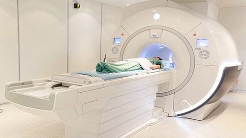
Máy chụp cộng hưởng
2.2 Preparation for implementation Performer: Specialist doctor, radiology technician and nurse; Vehicles: Resonant angiography machine of 1 Tesla or more; film, film printers and image storage systems; Medicines: Antiseptics for skin and mucous membranes; sedatives and contrast agents; Medical supplies: 10ml syringe, 18G intravenous catheter, distilled water (or physiological saline), cotton, gauze, gloves, sterile adhesive tape, medicine box and emergency equipment for drug accidents for patients. optical; Patient: Need to fast (no need to fast); be thoroughly explained about the procedure to coordinate with the doctor; sign a commitment to accept the procedure; check for contraindications; be instructed to change clothes of the magnetic resonance imaging room, remove contraindicated items; Licensed requires magnetic resonance imaging or complete medical records. 2.3 Steps of Magnetic Resonance Imaging of the Prostate Patient Position: Lie supine on the scanning table; the doctor selects and positions the signal receiver coil, then moves the table into the machine compartment and then locates the imaging area; The doctor placed an intravenous line for the patient with an 18G needle, connected to a double-bore electric injection pump (1 barrel containing magnetic contrast and 1 barrel containing physiological saline). The amount of contrast agent used for patients with the usual dose is 0.2ml/kg body weight; Location capture; Take before injecting magnetic contrast: Capture pulse sequences 1 - 2 - 3 - 4 with correct technique; Taken after injection of magnetic contrast agent; Perform magnetic contrast injection at a dose of 0.1 mmol/kg body weight at a rate of 2 mL/s; Capture the 5th and 6th pulse sequences correctly; Note: It is possible to use the receiver coil in the rectum or the receiver coil in the abdominal wall (in the case of thin patients) in the position on the abdomen in the pelvic region and then fixed with a lifeline, using the field. small observation.
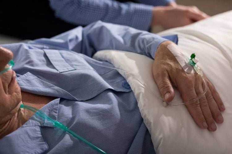
Sốc phản vệ là tai biến liên quan đến thuốc đối quang của chụp cộng hưởng từ tuyến tiền liệt
2.4 Evaluation of results On magnetic resonance imaging, the entire prostate gland and adjacent organs such as rectum, seminal vesicles, etc. must be clearly seen in the vertical, horizontal and horizontal directions; The doctor can assess the extent of the lesion's enhancement, if any; The doctor reads the medical condition, can give professional advice to the patient and the patient's family. 2.5 Complications and treatment After prostate magnetic resonance imaging, the patient may encounter the following conditions:
Fear, agitation: Doctors and family members should encourage and comfort the patient; Excessive fear, anxiety: The patient can be sedated under the supervision of an anesthesiologist; Complications related to contrast agents: Including nausea, urticaria, bronchospasm, laryngeal edema, hypotension, anaphylaxis, ... need to be handled according to the standard protocol. When performing magnetic resonance imaging of the prostate with magnetic contrast injection, patients need to strictly follow the doctor's instructions to obtain accurate diagnosis results and reduce the risk of complications.
Vinmec International General Hospital put into use the magnetic resonance imaging machine 3.0 Tesla Silent technology. Magnetic resonance imaging machine 3.0 Tesla with Silent technology of GE Healthcare (USA).
Silent technology is especially beneficial for patients who are children, the elderly, weak health patients and patients undergoing surgery. Limiting noise, creating comfort and reducing stress for customers during the shooting process, helping to capture better quality images and shorten the shooting time. Magnetic resonance imaging technology is the technology applied in the most popular and safest imaging method today because of its accuracy, non-invasiveness and non-X-ray use.
Master. Dr. Ton Nu Tra My is currently a Doctor of Radiology, Vinmec Central Park International General Hospital. Dr. Tra My used to be a lecturer in the Department of Diagnostic Imaging, Hue University of Medicine and Pharmacy and had a long time working at the Department of Diagnostic Imaging, Hue University of Medicine and Pharmacy Hospital.
To register for examination and treatment at Vinmec International General Hospital, customers can call Hotlines of hospitals or register online HERE.


