This is an automatically translated article.
The article was professionally consulted by Specialist Doctor II Khong Tien Dat, Doctor of Radiology - Department of Diagnostic Imaging - Vinmec Ha Long International General Hospital. The doctor has more than 14 years of experience in the field of diagnostic imaging.1. What is pleural ultrasound?
Pleural ultrasound is a technique capable of diagnosing diseases in the pleura effectively such as tumors, inflammation, lesions... In addition, pleural ultrasound is also highly appreciated in detecting pleural effusion , check the thickness of the pleura and tumor on the pleura, help guide the puncture, biopsy the pleura, drain the fluid.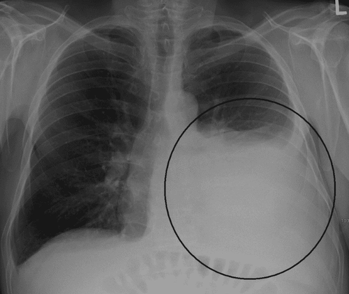
2. Pros and cons of pleural ultrasound
Pleural ultrasound is a modern technique of great value in supporting the examination, diagnosis and treatment of diseases. The outstanding advantages of the current pleural ultrasound technique:Flexible technique, can be performed at the hospital bed. The display image of pleural ultrasound is high resolution, fast and accurate. Ultrasonic equipment with compact size, can be moved when needed, convenient for the ultrasonic process. In particular, the pleural ultrasound technique does not have an irradiation dose, so it is safe and does not cause any damage to the organs when conducting the survey. The cost of implementation is reasonable and can be met with a wide range of patients. Besides the outstanding advantages mentioned above, pleural ultrasound also has certain disadvantages:
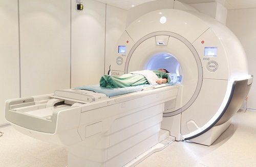
Quality of ultrasound images depends on ultrasound equipment at medical facilities. Ultrasound results are objective, requiring the sonographer to be highly qualified, professionally trained and experienced. Some pathological images are difficult to distinguish. It is necessary to combine with other techniques such as X-ray, magnetic resonance, ... to give the most accurate final conclusion.
3. Procedure for performing pleural ultrasound
3.1 Preparation. Patients undergoing pleural ultrasound do not need to prepare anything. Depending on the medical facility, the patient will need to change into a hospital uniform to facilitate the examination.Depending on the location to be examined and depending on the doctor's request, the patient can have the ultrasound in the lying or sitting position.
For the lying position: it is convenient to examine the pleura from the lateral and posterior costal angles of the diaphragm, the base of the lung parenchyma, and evaluate the diaphragm and pleura through the liver. For the upright sitting position: the transducer is placed in the intercostal spaces. With this position, the doctor can easily check for diseases of the chest wall, pleura, and find small amounts of pleural effusion. The ultrasonic device used has 2 probes:
Curved transducer with a frequency of 3.5 - 5 MHz. The flat probe has a frequency of 5 - 10 MHz. Proceed to apply sound conducting gel on the transducer position to obtain a clearer image.
3.2 Implementation steps Step 1: Move the transducer along the intercostal position. If there is a lesion, compare with the contralateral side and observe the change of respiratory rhythms.
Step 2: Observe the ultrasound image when the transducer moves to the position between the parietal pleura and visceral pleura, in case there is a negative space, this is a typical sign of pleural effusion. This step uses a 3.5 MHz probe to estimate the extent of the pleural effusion.
Step 3: With the case of pneumothorax, the ultrasound image will not show the sliding pleural image, the comet tail image and the enlarged pleural line.

4. Normal image on pleural ultrasound
Repeated echo signal When the sound waves meet the interface between the pleura and the air in the parenchyma, it will produce a repeated reverberation response showing straight lines parallel to the pleura. This is a normal image on pleural ultrasound. However, this imaging result is also seen in pneumothorax, so it is of little diagnostic value.
Comet tail sign This sign occurs when the sound waves encounter the atmosphere and the alveolar walls will show on the result screen successive responses in multiple directions.
Signs of sliding pleura The visceral and parietal leaves slide over each other, this sign will not appear when there is a pathology: pleural effusion, pneumothorax, pleural tumor. Therefore, this sign has high value in pleural ultrasound diagnosis.
In conclusion, the current pleural ultrasound technique plays a very important role in the diagnosis and treatment of diseases related to the pleura. Above are the advantages and disadvantages of the technique, hopefully providing readers with useful knowledge.
Please dial HOTLINE for more information or register for an appointment HERE. Download MyVinmec app to make appointments faster and to manage your bookings easily.


