This is an automatically translated article.
The article is professionally consulted by Master, Doctor Nguyen Thuc Vy - Radiologist - Department of Diagnostic Imaging - Vinmec Nha Trang International General Hospital.To evaluate the pulmonary ventilation status in some patients with chronic obstructive pulmonary diseases, bronchial asthma, obstructive bronchitis, ... Doctors often appoint magnetic resonance imaging when the lungs . The procedure of performing magnetic resonance ventilation of the lungs shows images of the chest, from which it is possible to evaluate lung function as well as pathologies.
1. Purpose of lung ventilation magnetic resonance imaging
Magnetic resonance ventilation of the lungs is a technique performed for the purpose of viewing images of the chest cavity, based on the imaging results to assess the ventilation function of the lungs.
The magnetic resonance imaging machine used is a machine with high magnetic force of 1.5T - 3T, using X129 gas or Helium-3 gas, with high polarization 3H-MRI.
2. Indications / contraindications for lung ventilation magnetic resonance imaging in which cases?
Magnetic resonance ventilation of the lungs is indicated in patients with bronchial asthma, obstructive bronchitis, chronic obstructive pulmonary disease, pulmonary fibrosis, ... to evaluate the ventilation function of the lungs. Pulmonary resonance imaging (MRI) is relatively contraindicated in patients with previous metal surgical clamps (over 6 months) or agoraphobia. Absolutely contraindicated in patients who use human electronic devices such as Neurostimulator, cochlear implant, automatic subcutaneous drug pumping device, pacemaker, anti-vibration machine, ...; or patients who have ever had vascular, intracranial, or orbital metal surgical clamps (less than 6 months), or have a serious illness requiring resuscitation equipment.
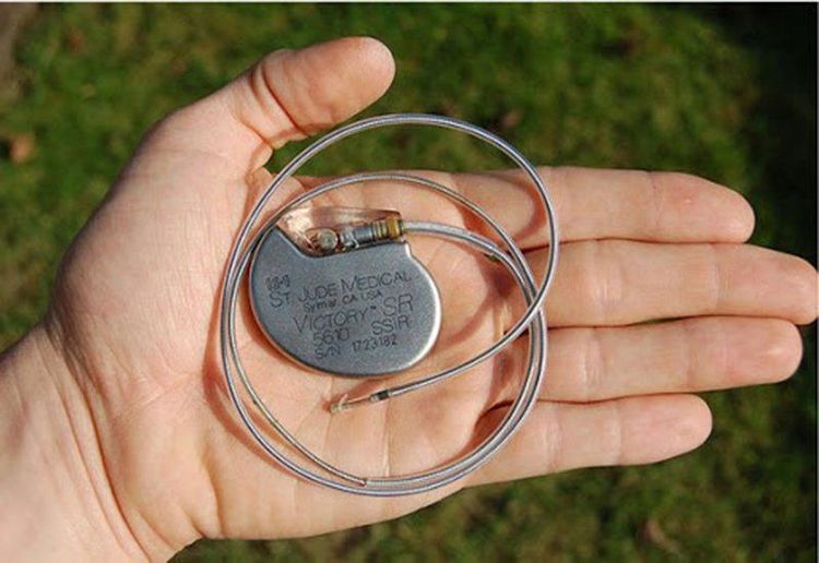
Bệnh nhân có thiết bị điện tử trên người không được chụp cộng hưởng từ
3. Procedure for performing pulmonary ventilation magnetic resonance imaging
For lung ventilation magnetic resonance imaging, the patient does not need to fast. However, it is important to check all contraindications prior to the scan.
The procedure to perform pulmonary ventilation magnetic resonance includes the following steps:
Step 1: Place the patient supine on the table, select and locate the signal receiver coil, then proceed to move the imaging table Enter the machine bay with the magnetic field and locate the area to capture. Step 2: Put the sandbag at the foot, place the coil to capture the whole chest, place the electrode to monitor the ECG. In case the patient has palpitations, heart palpitations, put Nitromint under the tongue, give the emergency call button and instruct the patient to use it. Step 3: Set the controller, proceed to capture the breathing rate and pay attention to the reduction of image noise during the magnetic resonance imaging of the lungs. Place the patient in a supine position in the direction of the injury if there is damage to the chest wall, in order to limit image noise due to breathing. Step 4: Capture the entire ribcage in the field from the base of the neck to the end of the diaphragm in 3 directions. Perform pulse sequence capture including: T2W pulse sequence - horizontal and cross-sectional imaging, image acquisition from the apex of the lung to the costophrenic angle, field of view (FOV) 380-400, phase difference from left to right (LR), the thickness of the cutting layer is 6 - 8 mm, the distance of 10-20% of the cut layer, if necessary, can be placed a magnetic barrier; T1W pulse sequence - cross-sectional imaging similar to the cross-sectional T2W pulse sequence. Step 5: Take a snapshot to assess lung ventilation by pulse sequences bSSFP, FLASH, HASTE, in 3 - 20 seconds, each inhalation - exhalation with a mixture of gases including Helium (Xenon), Nito, Oxygen and free gas (in pre-calculated proportions). Shoot in landscape, landscape and landscape orientation. The result is a color coded image. Assess the ventilation function of each lung area, calculate the ventilation ratio for each inhalation - exhalation. Step 6: The technician prints the film and transfers the obtained images to the doctor. Based on the images and parameters, the doctor will analyze and evaluate the ventilation level of the lungs and make a diagnosis.
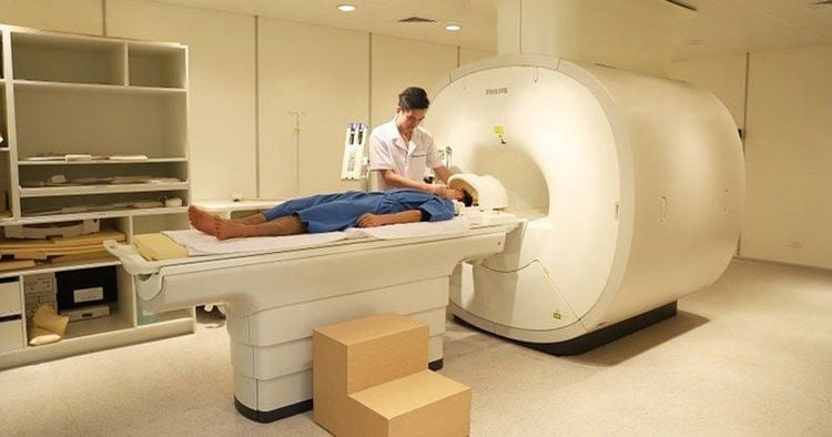
Quy trình thực hiện cộng hưởng từ thông khí phổi
Pulmonary magnetic resonance imaging is a technique that allows to evaluate the ventilation function of the lungs in some patients with lung diseases such as pulmonary fibrosis, chronic obstructive pulmonary disease, bronchial asthma, ...
To To meet the needs of medical examination, diagnosis and treatment, Vinmec International General Hospital has brought modern equipment and machines such as magnetic resonance imaging (MRI), computed tomography (CT), X- radiology, .. in medical examination and treatment, imaging diagnosis, lung cancer screening,... . In particular, the technicians who perform these techniques are a team of well-trained and qualified medical professionals who will give the most accurate image results combined with ultrasound results to provide a protocol. optimal treatment, minimizing dangerous complications for customers.
For detailed advice on this method, please come directly to Vinmec medical system or book online HERE.


