This is an automatically translated article.
Posted by CKI Doctor Vu Thi Hanh - Department of Diagnostic Imaging - Vinmec Hai Phong International General Hospital
The procedure for performing a wrist X-ray is quite simple, the results obtained will be of great help in diagnosing the disease and giving the appropriate treatment plan for the patient.
1. Concept of wrist X-ray
Wrist X-ray is a non-invasive imaging test that uses X-rays to reveal images of the bones of the wrist, thereby providing doctors with important information for diagnosing bone disease in the region. this.
2. Indications for X-ray of bones in the wrist region
The doctor will order an X-ray of the wrist in the following cases:
Diagnosis of fracture or dislocation Post-treatment examination (monitoring after fracture treatment, assessment of bone condition from which the surgeon can next treatment direction for the patient...). Look for lesions caused by infection or arthritis. Bone cancer lesions Locate the contrast medium in the bone or soft tissue around the wrist area.

Chụp X-quang cổ tay khi chẩn đoán tổn thương khớp
3. Procedure for performing a wrist X-ray
Step 1: Prepare the patient
An X-ray of the wrist requires almost no special preparation. For the patient who wears wrist jewelry, ask the patient to take it off. Ask the patient if she is pregnant or dangerous. chance to get pregnant. If it is really necessary to do X-ray testing to diagnose the disease, then measures must be taken to shield it to minimize the effect of X-rays on the fetus. Step 2: Prepare the media
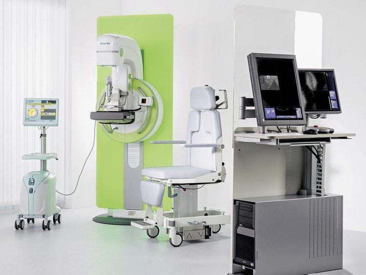
Digital X-ray machine Imaging table Step 3: Wrist X-ray procedure
X-rays are a form of radiation such as light or radio waves. It passes through most objects, including bodies. When it focuses on examining the wrist area, X-rays penetrate this area and are recorded as images on film. Different parts of the body absorb X-rays at different rates. Bones absorb most of the X-rays while soft tissues such as muscle, fat, and organs allow X-rays to pass through them. As a result, bone will appear white on film, soft tissue appears gray, and air is black. Most X-ray images are stored at the scanner. The stored images are easily accessible for disease diagnosis and disease monitoring.

Tia X đâm xuyên qua cổ tay và để lại hình ảnh trên phim
The technique is performed by a radiologist trained in wrist radiography. The patient is lying on the table or sitting next to the table, the patient's wrist will be placed on the table in 2 positions straight - tilted. In the straight wrist position: The wrist is placed prone on the imaging table, the central ray perpendicular to the film is located at the midpoint connecting the two rotator cuff and abutment processes. Note: In some cases: To clearly see the carpal bones, the central ray is receded 1cm below. If the central ray is inclined 30 degrees from the finger to the wrist, it is projected between the radial rotator cuff, then the shape of the joint with the bones of the wrist is very clear. In the slanted wrist position: Place the medial margin of the wrist (on the side of the ulna close to the film), align the wrist joint to the center of the film, adjust the junction between the rotator cuff and the ulna to be perpendicular to the film, the central ray square. The angle with the film is concentrated on the apex of the film, out the middle of the film. Some other techniques for arthroplasty: Pronation posture (back - front) Upright position (front - back) Tilt position Anterior tilt position (external rotation) Rotating upright position Rotating upright posture Fourth position anterior abduction (internal rotation) Straight anterior position with clear shinbone (Stecher position) During the scan, the patient must maintain the arm position that the medical staff has adjusted during the radiography process. After the scan has been completed the patient may be asked to wait for the radiologist to determine that all the necessary images have been revealed. The scan takes about 10 minutes.

Máy chụp X-quang tại bệnh viện ĐKQT Vinmec
4. Results of X-ray film of the wrist
Based on the X-ray film of the wrist, the radiologist views and reads the results:
Used to identify fractures and dislocations in trauma. Used to monitor the progress of bone healing in fracture trauma. Used to determine the actual bone age in children Used to diagnose osteomyelitis and bone cancer Used to determine the location of a contrast foreign object located in the bone or in the adjacent soft tissue. X-ray of the wrist is a simple technique, performed quickly and cheaply, helping to diagnose diseases of bones and joints in the wrist region, especially in trauma. However, today there are many other techniques such as MRI and ultrasound that can evaluate the coordination between bones, tendons-ligaments and soft tissues of the wrist.
Vinmec International General Hospital with a system of modern facilities, medical equipment and a team of experts and doctors with many years of experience in neurological examination and treatment, patients can completely peace of mind for examination and treatment at the Hospital.
To register for examination and treatment at Vinmec International General Hospital, you can contact Vinmec Health System nationwide, or register online HERE.
MORE
What is an X-ray: All you need to know Radiation levels when taking X-rays Everything you need to know about X-rays of the urinary system


