This is an automatically translated article.
The article is professionally consulted by Master, Doctor Nguyen Quang Duc - Doctor of Nuclear Medicine - Department of Diagnostic Imaging and Nuclear Medicine - Vinmec Times City International General Hospital.Radioisotopes have been widely used in the diagnosis and treatment of tumor pathologies. Creating images with radioisotopes can use scanners, gamma chambers or SPECT, PET machines, combined with CT scanners. Any device used to create images with radioisotopes has its own principles and advantages.
1. Using radioisotopes in tumor imaging
Currently, to diagnose and detect tumor pathologies, it is possible to use conventional imaging techniques such as ultrasound, X-ray, computed tomography or magnetic resonance, ...In addition, Nuclear medicine using radioisotopes is increasingly widely applied, as an imaging technique that allows imaging to detect tumors in many cancer diseases.
In which, tumor imaging by radioisotope has 3 main groups of methods:
Tumor imaging by contrast principle by gamma chamber, SPECT machine. This method includes imaging according to the principle of negative contrast and positive contrast. Tumor-specific imaging, also known as radioimmunoassay (Radio Immuno scintigraphy, abbreviated as RIS). Record the tumor according to the principle of metabolism by PET machine.
2. Imaging using radioisotopes
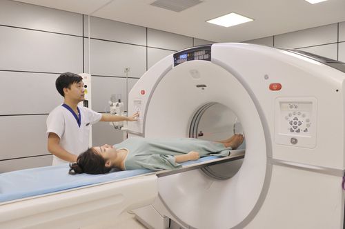
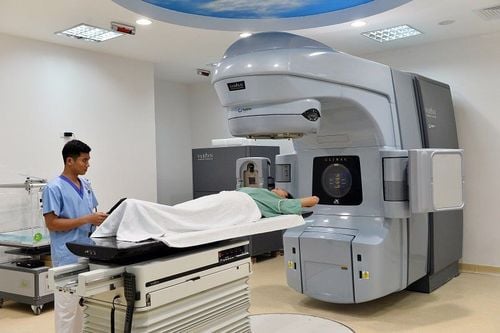
2.1 Radioisotope imaging with SPECT 2.1.1 Principle of radioisotope imaging with SPECT Radioisotope imaging with SPECT (short for Single Photon Emission Computed Tomography, tomography) single-photon irradiation) is a modern imaging technique that allows the detection of disease changes at the molecular level.The imaging technique of the SPECT machine is similar and developed from the CT-Scanner machine. The principle of imaging by radioisotope SPECT is as follows:
The radioactive source is inserted into the patient, to the location where the image needs to be taken orally or injected, ... to emit a beam of photon radiation. When the body emits a beam of radiation, the detector system rotates around the patient to record the radiation beam. Based on the physiological and pathological characteristics, the obtained images will show the concentrated radioisotope lesion area, thereby providing functional information of the examined organ and organ. The image obtained by SPECT machine is a 3-dimensional image, clearly displayed to serve to evaluate the function of some organs in the body, the process of cell metabolism.

Tạo ảnh bằng đồng vị phóng xạ
Probe and evaluate the body shape and function with high accuracy of some organs such as brain, thyroid, liver, bile, kidney, ... Diagnosis Diagnosis and early detection of brain lesions such as cerebral ischemia, cerebrovascular accident causing cerebral infarction, ... Diagnosis and evaluation of some cardiovascular diseases such as coronary artery disease, myocardial infarction, .. .. Using radioisotopes to scan thyroid, lungs, digestive system, liver, bile, kidneys, bones, lymphatic system. Using radioactive isotopes of iodine to identify neuroendocrine tumors.... In addition, the SPECT machine is also used to support biopsy samples or surgical resection. 2.1.3 Advantages of radioisotope imaging with SPECT machine With the above-mentioned principle of radioisotope imaging, SPECT is a modern diagnostic imaging device with the following advantages: early detection of tumors and metastases. Assess the possibility of recurrence compared with other methods such as ultrasound, computed tomography and magnetic resonance. Monitor response and evaluate effectiveness after treatment. Based on the results, the treating doctor will change the dose of chemotherapy or radiation therapy accordingly. 2.2 Radioisotope imaging with PET 2.2.1 Principle of radioisotope imaging with PET Radioisotope imaging with PET scanner (short for Positron Emission Tomography, tomography by single positron radiation) is a imaging technique at the cellular and molecular levels.
The principle of tumor imaging by PET machine is to apply the mechanism of concentrating radiopharmaceuticals selected specifically. To choose the appropriate radiopharmaceuticals, it is necessary to base on the physiological or metabolic differences between tumor tissue and normal tissue.
Principles of radioisotope imaging with PET machines are as follows:
Mark metabolites in tumors with a radioactive source that emits a beam of positron radiation. According to the circulation of blood, the radioactive source will concentrate in the tumor tissues and there participate in the metabolism, synthesis and transformation of cancer cells. Compared with the surrounding healthy tissues, the tumor-injured area will concentrate radiopharmaceuticals and clearly show the difference in radioactivity. Radioisotope imaging with PET machines will obtain functional images of specific cancer tissues from a very early stage, when cancer cells are still in the metabolic disorder stage. Meanwhile, diagnostic imaging by CT or MRI, ... only allows detection when the cancer organization is destroyed to a large extent. 2.2.2 Focusing mechanism using radioactive isotopes to create images of PET machine The basic principle to obtain tumor images of PET machine is to apply the mechanism of concentrating radiopharmaceuticals in cells. cancer . This mechanism is used as follows:
Compared with normal tissues, tumors have a very fast growth rate, thus increasing the utilization rate of DNA precursors in the tumor. Using the radioisotope C-11 conjugated to these progenitors will image areas where malignant lesions are concentrated and show areas of increased radioactivity on imaging. Compared with normal tissues, tumors have a higher rate of protein synthesis, thus increasing the process of using, transporting and combining a variety of amino acids in cancer organizations. Using the radioisotope C-11 bound to these amino acids will image areas where malignant lesions are concentrated and show areas of increased radioactivity on the images. Compared with normal tissues, tumors have more anaerobic and aerobic glucose breakdown, so the demand for glucose by the tumor is higher. The attached radioisotope 18F-FDG or C-11 will capture the areas where the malignant lesion is concentrated and show the area of increased radioactivity on imaging. There are many radiopharmaceuticals used to image tumors, depending on the characteristics and properties of cancer cells, the appropriate radiopharmaceuticals will be used for imaging.

Tạo ảnh bằng đồng vị phóng xạ với máy PET
Recording myocardial perfusion, brain perfusion, ... Detecting cancerous tumors. Monitor and evaluate the results of treatment. 2.2.4 Advantages of radioisotope imaging with PET machine With the principle of radioisotope imaging and the principle of concentration of radiopharmaceuticals mentioned above, PET imaging has the following advantages: :
Provides both anatomical and metabolic functional images of the tumor, whereas CT and MRT diagnostics provide only anatomical images. Has higher sensitivity and specificity, helping to detect cancer at an early stage. Early and accurate assessment of cancer response. Distinguishing between cancerous tissue and fibrous or necrotic scar tissue, ... Radioisotope imaging with SPECT and PET machines in combination with CT scanning are modern imaging methods to help detect detect cancer at an early stage, and monitor and evaluate the response during and after treatment.
Currently, Vinmec International General Hospital has become a leading prestigious address in disease screening with modern techniques, proud to be the first private hospital in Vietnam to deploy this technique. Modern and accurate diagnosis with the most modern 128-sequence PET/CT system in Southeast Asia, for accurate images and short imaging time, the team of doctors are leading experts with high expertise and rich experience. Experience creates trust for customers when experiencing medical examination and treatment services at Vinmec.
If you have a need for consultation and examination at Vinmec Hospitals under the nationwide health system, please book an appointment on the website for service.
Please dial HOTLINE for more information or register for an appointment HERE. Download MyVinmec app to make appointments faster and to manage your bookings easily.